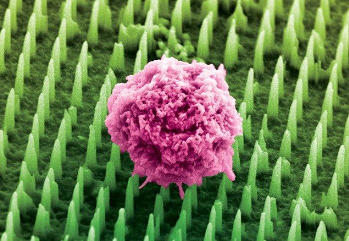
![Colourised image of Nanoneedles [Chippani/King's College London]](https://www.genengnews.com/wp-content/uploads/2025/06/low-res-7.jpeg)
Colorized image of Nanoneedles [Chippani/King’s College London]
Scientists at King’s College London have developed a patch comprising tens of millions of microscopic nanoneedles, which they suggest could offer a painless and less invasive alternative to biopsies for millions of patients worldwide who undergo these procedures each year to detect and monitor diseases such as cancer or Alzheimer’s.
The nanoneedle patch collects molecular information directly from tissues without damaging them, and because the porous silicon nanoneedles are 1,000 times thinner than a human hair and do not remove tissue, they also cause no pain. The technology could feasibly allow healthcare teams to monitor disease in real time and perform multiple, repeatable tests from the same area—something impossible with standard biopsies. For patients this could mean earlier diagnosis and more regular monitoring, transforming how diseases are tracked and treated.
Research lead Ciro Chiappini, PhD, Senior Lecturer in Nanomaterials and Biointerfaces at King’s College London, said, “We have been working on nanoneedles for twelve years, but this is our most exciting development yet. It opens a world of possibilities for people with brain cancer, Alzheimer’s, and for advancing personalized medicine. It will allow scientists— and eventually clinicians—to study disease in real time like never before.”
Senior and corresponding author Chiappini, and colleagues reported in Nature Nanotechnology on use of the nanoneedle patch technology applied to brain cancer tissue from human biopsies and mouse models. In their paper, titled “Nanoneedles enable spatiotemporal lipidomics of living tissues,” the team concluded, “This approach establishes a minimally perturbative method for spatiotemporal lipidomics of live tissues, with transformative potential for studying tissue dynamics and therapeutic responses.”
Spatial biology provides high-content diagnostic information by mapping the molecular composition of tissues, the team wrote. “In particular, spatial lipidomics gives insight into metabolism and its perturbations during physiological and pathological processes,” they explained. “However, spatial biology typically relies on destructive processes in non-living tissues, limiting its applicability for the study of unique samples or to track the spatiotemporal dynamics of living systems.”
Biopsies are among the most common diagnostic procedures worldwide, performed millions of times every year to detect diseases. However, they are invasive, can cause pain and complications, and can deter patients from seeking early diagnosis or follow-up tests. Traditional biopsies also remove small pieces of tissue, limiting how often and how comprehensively doctors can analyze diseased organs like the brain.
In contrast, vertical nanoprobes, such as nanoneedles, could allow repeated sampling from the same live specimen. “Nanoprobes can access intracellular compartments to collect biomolecules with minimal disruption to cell function, enabling repeated or continuous sampling,” the scientists suggested. “Among them, porous silicon nanoneedles are particularly adept to achieve this vision.”
The team’s newly published paper describes development and preclinical evaluation of a patch covered in tens of millions of these nanoneedles. In preclinical studies, the team applied the patch to brain cancer tissue taken from human biopsies and from mouse models. The nanoneedles extracted molecular fingerprints—including lipids, proteins, and mRNAs—from cells, without removing or harming the tissue.
The tissue imprint is then analyzed using mass spectrometry and artificial intelligence, giving healthcare teams detailed insights into whether a tumor is present, how it is responding to treatment, and how disease is progressing at the cellular level. “Using nanoneedles, we repeatedly generated non-destructive imprints of brain tissue, called molecular replicas,” the scientists further explained. “These replicas well preserved the relative composition and spatial distribution of the original samples, allowing accurate mapping of brain regions and their lipid profiles. This approach enabled effective disease grade prediction, matching the performance of direct tissue analysis.”
Chiappini added, “This approach provides multidimensional molecular information from different types of cells within the same tissue. Traditional biopsies simply cannot do that. And because the process does not destroy the tissue, we can sample the same tissue multiple times, which was previously impossible.”
The researchers suggest that technology could be used during brain surgery to help surgeons make faster, more precise decisions. For example, by applying the patch to a suspicious area, results could be obtained within 20 minutes and guide real-time decisions about removing cancerous tissue. “By preserving lipid spatial distribution and enabling non-destructive, repeated sampling, nanoneedles offer a powerful platform for spatiotemporal, omics-level molecular diagnostics,” the team stated.
Made using the same manufacturing techniques as computer chips, the nanoneedles can be integrated into common medical devices such as bandages, endoscopes and contact lenses. “Their integration into medical devices enables the analysis of accessible tissues, including intraoperative sampling during glioma surgery and longitudinal monitoring of mucosal surfaces,” the authors continued.
Dr Chippani added, “This could be the beginning of the end for painful biopsies. Our technology opens up new ways to diagnose and monitor disease safely and painlessly—helping doctors and patients make better, faster decisions.”
The breakthrough was possible through close collaboration across nanoengineering, clinical oncology, cell biology, and artificial intelligence—each field bringing essential tools and perspectives that, together, unlocked a new approach to non-invasive diagnostics.