
Credit: Getty/ BeholdingEye
A sterility test for cell therapy products returns yes/no results within 30 minutes for a range of microbial contaminants. Such detection speed is the result of combining machine learning (ML) and ultraviolet (UV) absorbance spectroscopy to detect microbial contamination in cell therapy products.
Because ML-enabled UV absorbance spectroscopy is noninvasive, no cells are required for testing, so the cell product isn’t compromised. Because it doesn’t require additional incubation periods and growth enrichment mediums, this method is significantly faster than the approximately 14 days required by the testing methods outlined in United States Pharmacopeia chapter 71, as well as the approximately seven days required by advanced, rapid microbiological methods.
Continuous monitoring potential
This proof-of-principle research has the potential to become a preliminary test during cell therapy manufacturing to provide real-time, continuous culture monitoring for microbial contamination detection.
In addition to speed, ML-aided UV absorbance spectroscopy is label-free and uses only a small volume—less than 1 mL—of cell culture supernatant. The ML algorithm can be trained using relatively small quantities of data.
This ML-enabled sterility testing method was detailed in a recent paper by Shruthi Pandi Chelvam, first author and senior research engineer at the Critical Analytics for Manufacturing Personalized-Medicine (CAMP) interdisciplinary research group within the Singapore-MIT Alliance for Research and Technology (SMART); Rajeev Ram, PhD, principal investigator at SMART CAMP and professor at the Massachusetts Institute of Technology (MIT); and researchers at MIT, A*STAR Skin Research Labs, and the National University of Singapore.
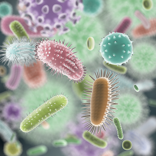
For the tests, Chelvam and colleagues spiked mesenchymal stem cells (MSCs) with seven microbial organisms—S. aureus ATCC 6538, P. paraeruginosa ATCC 9027, B. spizizenii ATCC 6633, C. sporogenes ATCC 19404, C. albicans ATCC 10231, E. coli K-12 (ATCC 25404), and C. acnes ATCC 6919—and detected contamination at inocula as low as 10 colony-forming units (CFUs).
Analysis of 418 samples showed a mean true positive of 92.7% and a mean true negative of 77.7%. Excluding an anomalous sample from a patient who exhibited very high nicotinic acid levels improved the true negative rate to 92%. Contaminant detection, the scientists concluded, seems to depend on detecting the spectral differences between nicotinic acid and nicotinamide metabolites.
When the MSCs were inoculated with 10 CFUs of E. coli at the 0-hour time point as part of time-to-detection experiments, contamination was detected approximately 21 hours after the cells were exposed to the bacteria. “That is comparable to the performance of USP 71 testing methods,” Ram and Chelvam told GEN. “However, ML-enabled UV spectroscopy took longer to detect contamination compared to rapid microbiological methods, indicating lower sensitivity.”
This method, they say, “was designed to be implemented as part of the cell therapy manufacturing process, as a form of preliminary testing.” If the sample is contaminated, they recommend using regulatory-approved single time-point confirmation testing.
“Time to detection cannot be generalized using E. coli only,” the scientists cautioned. Therefore, they plan to extend the model to “cell types that are widely used in cell therapy manufacturing,” and to develop “testing to confirm contamination detection in other strains of bacteria and in additional microorganisms, such as yeast and fungi,” Ram and Chelvam said.