
In a groundbreaking development poised to transform the landscape of surgical oncology, a team of researchers has unveiled the first clinical trial demonstrating the use of nerve-specific fluorescence during head and neck surgeries. The Phase 1 trial, recently published in Nature Communications, showcases the remarkable precision and potential safety benefits of this innovative imaging technique designed to improve nerve visualization intraoperatively. This approach promises to drastically reduce the risk of nerve damage, a notorious complication in surgeries where vital nerve structures lie in close proximity to malignant tissues.
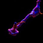
Nerve injury during head and neck surgery is a significant clinical challenge, often resulting in debilitating functional deficits including loss of sensation, motor control, or even swallowing and speech impairments. Traditionally, surgeons have relied heavily on anatomical knowledge and intraoperative experience to identify and preserve these nerves, but this method has inherent limitations. The advent of nerve-specific fluorescent dyes represents a revolutionary technological leap, allowing surgeons to literally “see” nerves glowing in real-time under near-infrared light conditions during the procedure.
This Phase 1 clinical trial led by Lee, YJ., Orosco, R.K., Bouvet, M., and their team focused on evaluating the safety and feasibility of administering novel fluorescent agents specifically conjugated to nerve tissue markers. These agents selectively bind to peripheral nerves and emit a distinctive fluorescence, thus enhancing nerve delineation against the complex background of surrounding tissues. The trial enrolled patients undergoing various head and neck surgical interventions where nerve preservation is critical, enabling real-time assessment of the fluorescent imaging’s accuracy.
.adsslot_FNjM4coKX9{width:728px !important;height:90px !important;}
@media(max-width:1199px){ .adsslot_FNjM4coKX9{width:468px !important;height:60px !important;}
}
@media(max-width:767px){ .adsslot_FNjM4coKX9{width:320px !important;height:50px !important;}
}
ADVERTISEMENT
Technical nuances of the fluorescent agents used include their biocompatibility, rapid systemic clearance, and strong fluorescent signal in the near-infrared spectrum around 700–900 nm wavelengths. These properties are vital, as they avoid interference from native tissue autofluorescence and allow deeper tissue penetration of the excitation light, crucial for visualizing nerves that lie beneath multiple tissue layers. Importantly, the dyes exhibit minimal off-target binding, reducing false positives and enhancing the surgeon’s confidence in distinguishing nerve fibers from blood vessels and connective tissues.
The implications of this nerve-specific fluorescence technology extend well beyond mere visualization. By providing high-contrast images of nerve anatomy, these dyes enable more precise dissection, reducing inadvertent nerve transection or stretching. Enhanced nerve identification may result in shorter operative times and decrease the need for additional corrective surgeries. Moreover, better nerve preservation correlates strongly with improved postoperative quality of life, including preserved speech, swallowing function, and facial expressions, which are paramount to patient well-being after head and neck cancer surgeries.
Throughout the trial, patients tolerated the fluorescent agents without significant adverse effects, underscoring the biocompatibility of the compounds. Imaging sessions were seamlessly integrated into the surgical workflow, demonstrating practicality in real-world operative settings. Surgeons reported that the augmented imaging fundamentally enhanced their ability to navigate complex anatomical regions, especially around the parotid gland, skull base, and cranial nerve branches. The real-time feedback allowed dynamic adjustments during dissection, setting a new standard in surgical precision.
This approach also opens avenues for training and education, as the visualization of nerves in fluorescence mode could serve as an invaluable teaching tool for surgical trainees. It democratizes expert knowledge by making intricate nerve anatomy visually explicit rather than purely conceptual, potentially accelerating the acquisition of surgical skill and reducing the learning curve for complex head and neck procedures.
Given the Phase 1 design, the trial primarily established safety parameters and proof-of-concept efficacy rather than long-term functional outcomes. Future studies with larger cohorts and longer follow-up periods will be essential to quantify functional improvements in nerve preservation and validate the technology’s superiority over existing intraoperative nerve monitoring techniques. Researchers are optimistic about combining fluorescence imaging with other modalities like electrophysiological monitoring to create an integrated, multimodal nerve-sparing surgical platform.
The technology itself is the culmination of interdisciplinary innovation involving molecular chemistry, biomedical optics, and surgical science. The fluorescent nerve markers were painstakingly engineered to target myelin-associated glycoproteins and other nerve-specific epitopes with high affinity. The synthesis protocols ensure stability in vivo and compatibility with existing surgical imaging devices, facilitating rapid translation from bench to bedside. This cross-disciplinary collaboration epitomizes the future trajectory of personalized and precision surgery.
Importantly, the trial’s results underscore the feasibility of bringing such advanced imaging technology into routine clinical care. If further validated, this modality could become a standard adjunct in head and neck, neurosurgical, and even peripheral nerve surgeries, where detailed nerve visualization is crucial. It represents a paradigm shift from conventional ‘visible light’ surgery to fluorescence-guided interventions that enhance the surgeon’s visual acuity beyond the capabilities of the naked eye.
In a broader context, the success of nerve-specific fluorescence imaging may catalyze similar approaches targeting other critical anatomical structures such as lymphatic channels, blood vessels, and tumor margins. Multiplexed fluorescence probes with distinct spectral profiles could soon allow simultaneous visualization of multiple tissue types, further improving surgical outcomes and reducing complications.
Commercially, this technology may induce a renaissance in surgical imaging equipment development, with industry players racing to integrate nerve-specific fluorescence capabilities into existing surgical endoscopes and microscopes. Accessibility and cost-effectiveness considerations will be vital to ensure widespread adoption, especially given the global burden of head and neck cancers concentrated in resource-limited settings.
Ethically, the use of fluorescent contrast agents raises questions about safety, long-term biodistribution, and potential immunogenic responses. Thus, rigorous regulatory scrutiny and post-market surveillance will be necessary to monitor rare adverse events and ensure patient safety. The transparency of the ongoing research process, including data sharing and cross-institutional collaborations, will be key in establishing clinical confidence and public trust.
Ultimately, the Phase 1 trial by Lee and colleagues marks a milestone in surgical innovation. By transforming the invisible into the visible, nerve-specific fluorescence imaging empowers surgeons with unprecedented anatomical clarity. It signifies a hopeful future where nerve injury risks diminish and functional preservation becomes the norm rather than exception in complex head and neck surgeries.
As surgical science marches toward an era of molecular-guided precision, the optics revolution spearheaded by this study heralds new vistas of patient-centric care. The convergence of chemistry, optics, and clinical expertise epitomizes the transformative potential of interdisciplinary collaboration to solve some of medicine’s most daunting challenges.
This pioneering study lays the foundation for a new standard in surgical practice, reminding us how illuminating the unseen can fundamentally change outcomes, not only in the operating room but in the lives afterward.
Subject of Research: Intraoperative nerve-specific fluorescence visualization for enhanced nerve identification during head and neck surgery
Article Title: Intraoperative nerve-specific fluorescence visualization in head and neck surgery: a Phase 1 trial
Article References:
Lee, YJ., Orosco, R.K., Bouvet, M. et al. Intraoperative nerve-specific fluorescence visualization in head and neck surgery: a Phase 1 trial. Nat Commun 16, 6060 (2025). https://doi.org/10.1038/s41467-025-60737-x
Image Credits: AI Generated
Tags: clinical trial nerve imagingfluorescent dyes in surgeryhead and neck surgery innovationsintraoperative nerve visualization techniquesnear-infrared light imagingnerve injury challenges in surgerynerve-specific fluorescencepreserving nerve function during operationsreducing nerve damage in surgerysafety benefits of nerve imagingsurgical oncology advancementstechnological advancements in surgical techniques