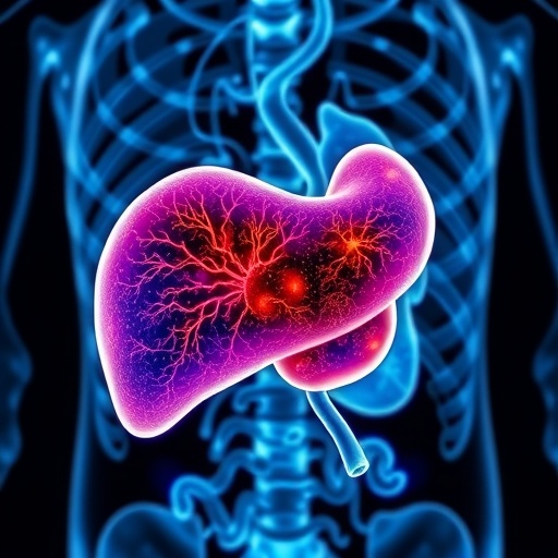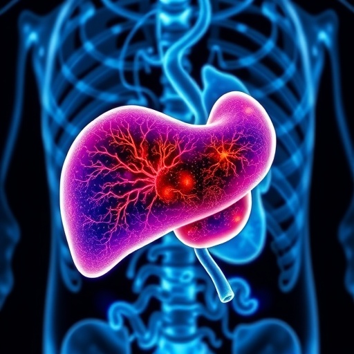
In an innovative study that stands to revolutionize the treatment of liver tumors, researchers have introduced a novel framework known as the RFiLM U-Net. This cutting-edge approach amalgamates radiomic features with linear modulation techniques, leading to unparalleled advancements in liver tumor segmentation. The study, conducted by a team of experts including Tsai, Agrawal, and Dash, focuses on enhancing precision in medical imaging, thus facilitating better clinical outcomes for patients diagnosed with liver malignancies.
The significance of accurate liver tumor segmentation cannot be overstated. Precise delineation of tumors is crucial for effective treatment planning, which often includes surgical resection, radiation therapy, or transarterial chemoembolization. Traditional methods of tumor segmentation often fall short in terms of accuracy and reliability, leaving a critical gap that RFiLM U-Net aspires to fill. This new model leverages advanced machine learning techniques to deliver comprehensive insights that are essential in the context of personalized medicine.
At the core of the RFiLM U-Net is the integration of radiomic features, which pertain to quantitative data extracted from medical images. Radiomics is an emerging field that utilizes high-throughput methods to decode the phenotypic characteristics of tumors, thereby providing valuable prognostic and predictive information. By harnessing these features, the RFiLM U-Net aims to enhance the segmentation process, allowing for a more effective analysis of tumor morphology and heterogeneity.
The underlying architecture of the RFiLM U-Net employs a unique linear modulation approach that refines the images used for tumor identification. This model not only processes the visual data more effectively but also aids in minimizing uncertainty, which is a common challenge faced in imaging diagnostics. The innovative structure functions by modulating the information flow within the neural network, leading to more robust feature representations, ultimately translating to improved segmentation accuracy.
One of the study’s pivotal aspects is its validation phase, where the researchers tested the RFiLM U-Net against conventional segmentation models. The results demonstrated that the new framework significantly outperformed existing methodologies in terms of both segmentation accuracy and computational efficiency. Such a leap in performance reflects the promising future of integrating advanced artificial intelligence techniques into the domain of medical imaging.
Furthermore, the researchers highlighted the model’s ability to generalize across various imaging modalities, including CT and MRI scans. This versatility is of paramount importance as it suggests that the RFiLM U-Net could be deployed in a wide array of clinical settings, ultimately benefiting a larger patient population. The adaptability of this model underscores the potential for wider application in oncological practices aimed at improving patient care.
The clinical implications of this research extend far beyond mere segmentation enhancements. The increased accuracy in tumor delineation fosters better treatment planning, thereby improving prognostic outcomes for patients. This could lead to more tailored therapeutic approaches, where interventions are closely aligned with the specific tumor characteristics discerned through advanced imaging techniques.
Moreover, the RFiLM U-Net model facilitates a more thorough assessment of tumor response to treatment over time. By employing this model in longitudinal studies, clinicians can better track the effectiveness of various therapeutic strategies based on real-time evaluation of tumor dynamics. This could serve as a game-changer in the field of oncology, leading to more effective interventions and improved quality of life for patients.
As the research community continues to explore the extensive landscape of artificial intelligence in medicine, studies such as these are indicative of the bright prospects that lie ahead. By refining methodologies for tumor segmentation, scientists can pave the way for smart technologies that enhance decision-making in diagnostics and treatment. The RFiLM U-Net illustrates this potential, shining a light on how integration of computing power and medical expertise can yield significant advancements in health outcomes.
The successful application of the RFiLM U-Net raises a pertinent question about the future of medical imaging. As clinicians, researchers, and technologists collaborate more closely, the possibilities for advancement become virtually limitless. The innovation showcased in this study may spur additional research aimed at further integrating AI-driven solutions into medical practices, ultimately leading to a paradigm shift in how tumors are diagnosed and treated.
In conclusion, the development of the RFiLM U-Net marks a significant milestone in the field of liver tumor segmentation, combining the strengths of radiomics and machine learning to achieve outstanding performance. By prioritizing precision and adaptivity, this framework offers immense promise for enhancing clinical practices and patient care. As such, it stands as an exemplary model for future research endeavors focused on integrating artificial intelligence in medicine, where the confluence of technology and healthcare continues to forge new paths toward improving patient outcomes.
The work represented in this innovative study not only contributes to the academic field but also carries the potential to dramatically transform clinical practices in oncology. As we stand on the brink of a new era in medical imaging, it is imperative that we continue to foster and support such initiative-driven research to fully realize the capabilities of modern technology in enhancing healthcare delivery.
Subject of Research: Radiomic Feature-Integrated Linear Modulation Network for Precise Liver Tumor Segmentation
Article Title: RFiLM U-Net: Radiomic Feature-Integrated Linear Modulation Network for Precise Liver Tumor Segmentation
Article References:
Tsai, LW., Agrawal, A., Dash, P. et al. RFiLM U-Net: Radiomic Feature-Integrated Linear Modulation Network for Precise Liver Tumor Segmentation.
J. Med. Biol. Eng. 45, 177–186 (2025). https://doi.org/10.1007/s40846-025-00938-3
Image Credits: AI Generated
DOI: https://doi.org/10.1007/s40846-025-00938-3
Keywords: Liver Tumor, RFiLM U-Net, Radiomics, Machine Learning, Medical Imaging, Tumor Segmentation.
Tags: advanced medical imaging techniquesinnovative cancer treatment methodologiesliver tumor segmentationmachine learning in healthcarepersonalized medicine applicationsprecision in liver cancer treatmentquantitative imaging analysisradiomic features in oncologyradiomics in tumor characterizationRFiLM U-Net frameworksurgical planning for liver tumorstumor delineation accuracy


