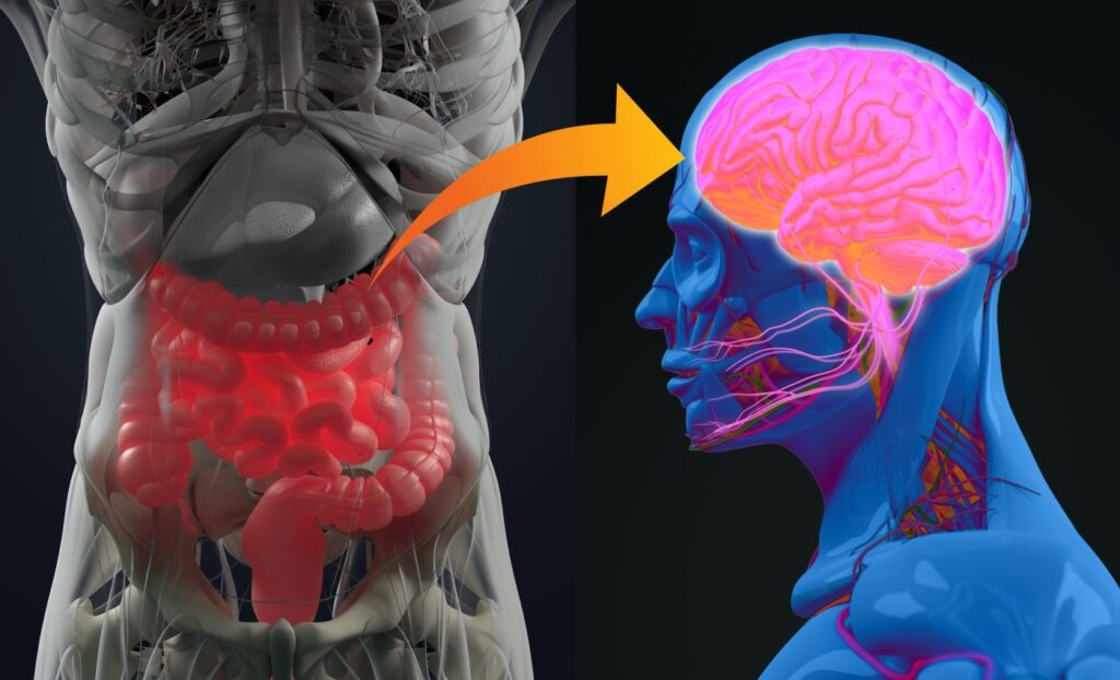
Alzheimer’s disease is a neurodegenerative disorder characterized by brain alteration including synaptic loss, chronic inflammation, and neuronal cell death. Scientists have found evidence that the gut and the brain communicate through the neurons placed in both organs. Recent studies have explored how disruptions or abnormalities in the gut microbiome could raise the risk of neurological diseases. Now, scientists led by the Institute of Nanotechnology in Italy, in collaboration with the European Synchrotron Radiation Facility (ESRF) in France have discovered how X-ray micro- and nano-tomography can provide clues on the processes that link the gut neurons with those in the brain and may trigger Alzheimer’s.
The findings are published in Science Advances in an article titled, “Investigating gut alterations in Alzheimer’s disease: In-depth analysis with micro- and nano-3D X-ray phase contrast tomography.”
“Alzheimer’s disease (AD), a debilitating neurodegenerative disorder, remains one of the foremost public health challenges affecting more than 30 million people worldwide with the etiology still largely enigmatic,” the researchers noted. “The intricate gut-brain axis, serving as a vital communication network between the gut and the brain, appears to wield influence in the progression of AD. Our study showcases the remarkable precision of x-ray phase-contrast tomography (XPCT) in conducting an advanced three-dimensional examination of gut cellular composition and structure.”
The gut microbiota, which refers to the microorganisms in the intestinal tract, plays a key role in human health and influences brain function, cognition, and behavior. “There are already many studies that support that changes in the gut composition can contribute to Alzheimer’s onset and progression,” explained Alessia Cedola, PhD, a researcher from the Institute of Nanotechnology in Italy and corresponding author of the article.
In particular, dysbiosis, which is the process by which there is a loss of microbial diversity, induces the prevalence of dangerous bacteria producing toxic metabolites promoting inflammation, and, consequently, the breakage of the gut/brain barriers.
The scientists sought to determine how exactly happens when gut dysbiosis occurs.
“The main hypothesis is that changes trigger the escape of bad bacteria from the gut, entering the circulation, reaching the brain, and triggering Alzheimer’s, but evidence is still poor,” added Cedola.
The scientists discovered that nano- and micro-XPCT is a powerful tool that could be used to study structural and morphological alterations in the gut, without tissue manipulation. The team came to the ESRF to scan samples on beamline ID16A. “Thanks to this technique we can image soft biological tissues with excellent sensitivity in 3D, with minimal sample preparation and without contrast agents,” explained Peter Cloetens, scientist in charge of ID16A and co-author of the publication.

The data from their experiments showed the changes in cell abundance and organization in the tissues, as well as structural alteration in different tissues of mice affected with Alzheimer’s. Specifically, it showed relevant alterations in the villi and crypts of the gut, cellular transformations in Paneth and goblet cells, along with the detection of telocytes, neurons, erythrocytes, and mucus secretion by goblet cells within the gut cavity. All these elements, when working correctly, maintain gut health, support digestion, and protect the intestinal lining from damage.
“This technique represents a real breakthrough for the thorough analysis of the gut, and it could be pivotal in early detection and prognosis of the disease,” said Cedola. She added: “As a long-time user of the ESRF, I can attest to the incredible opportunities that this facility provides for cutting-edge research, and the nanoimaging beamline, especially with the EBS. Coming to the ESRF has been instrumental in advancing our understanding of the gut-brain axis in Alzheimer’s disease.”
Looking toward the future, the scientists plan to further exploit the capabilities of XPCT to study how the gut communicates with the central nervous system. The team aims to investigate the enteric nervous system and its role in Alzheimer’s disease.
“By gaining a deeper understanding of these processes, we hope to identify new therapeutic targets and develop innovative treatments for this devastating disease. The ESRF will undoubtedly continue to play a crucial role in our research, and we look forward to many more exciting discoveries in the years to come,” concluded Cedola.