In a groundbreaking advancement at the intersection of forensic science and medical imaging, researchers have unveiled a novel approach to forensic age estimation leveraging the precision of elbow magnetic resonance imaging (MRI) combined with sophisticated data mining techniques. The study, conducted within a Chinese population, offers a significant leap forward in the quest for accurate legal age classification, a matter of critical importance in judicial and administrative processes worldwide. This pioneering research equips forensic experts with a more reliable toolset for determining chronological age, thereby enhancing the integrity of age-related legal decisions.
Traditional forensic age estimation methods usually rely on physical examinations, dental assessments, or analysis of hand-wrist radiographs. However, these approaches are often subject to variability due to ethnic differences, environmental conditions, and individual biological uniqueness. The innovative use of elbow MRI scans addresses some of these challenges by providing high-resolution images of ossification centers and cartilage that show characteristic developmental stages at different chronological ages. This imaging modality is non-invasive and free from radiation exposure, rendering it highly suitable for repeated forensic evaluation.
Furthermore, the incorporation of advanced data mining algorithms into the classification process introduces a powerful dimension of objectivity and analytic rigor. Data mining enables the extraction of complex patterns across the image datasets, uncovering subtle markers that might elude conventional visual inspection. By training these algorithms on a robust dataset drawn from a Chinese cohort, the research authenticates age estimation models tailored to population-specific developmental patterns, thereby refining accuracy and minimizing error margins.
Age thresholds are pivotal in numerous legal contexts, including criminal responsibility, consent to medical treatment, and eligibility for social services. Erroneous age estimation can lead to unfair penalties or denial of rights, underscoring the ethical and legal imperatives of methodological precision. The research team’s focus on legal age threshold classification using elbow MRI not only advances forensic science but also addresses a societal demand for enhanced fairness and transparency in age-related adjudications.
Another compelling aspect of this study is the comprehensive characterization of the ossification stages observable in MRI scans of the elbow joint. The investigation delineates specific morphological and structural markers corresponding to distinct developmental phases. Such detailed morphometric analyses empower forensic practitioners to anchor age estimations in objective anatomical landmarks rather than relying solely on subjective interpretation, a critical improvement in forensic evidence evaluation.
Integrating machine learning frameworks with medical imaging data allows continuous algorithmic adaptation as more data becomes available, fostering an evolving and self-improving system. This dynamic methodology stands in contrast to static reference tables, which are prone to outdatedness and do not account for inter-individual variability. The fusion of big data analytics with precise imaging heralds a future whereby forensic age estimation could achieve unprecedented levels of specificity and reliability.
From a technical standpoint, the MRI protocols employed in this study prioritize sequences optimized for cartilage and bone visualization, ensuring that the key developmental indicators are clearly discernible. This meticulous imaging technique undergirds the subsequent data mining process, ensuring that input quality is maintained at the highest standard. The resultant data integrity amplifies the confidence levels associated with age classifications derived from this approach.
Notably, this research traverse beyond mere age estimation, opening avenues for the application of similar methodologies to other anatomical regions or diverse demographic cohorts globally. The modular design of the analytic framework lends itself to adaptability, making it a versatile blueprint for subsequent forensic advancements. Cross-cultural and cross-ethnic validation studies could further expand the utility and generalizability of these findings.
The ethical dimension of forensic imaging and age estimation is explicitly acknowledged in this pioneering work. By reducing reliance on invasive methods and enhancing objective data analysis, the approach respects individual rights while reinforcing societal protection mechanisms. It embodies an ideal balance between forensic necessity and humanitarian consideration, pushing the discipline towards more ethical and scientifically grounded practices.
Forensic age estimation has also encountered challenges in juvenile identification, especially in the contexts of immigration and human trafficking where age documentation is often unreliable or missing. The precise, scientifically verifiable age estimation tools demonstrated in this study could significantly influence how authorities verify ages in such sensitive cases, ensuring that minors receive age-appropriate protections and assistance.
Moreover, the study’s integration of forensic science with cutting-edge medical technology epitomizes interdisciplinary innovation, a trend that continues to reshape modern science. By marrying radiologic imaging with computational intelligence, this approach exemplifies how traditional forensic questions can find solutions in the rapidly evolving landscape of digital and biomedical technologies, signaling a paradigm shift for decades to come.
The dataset underpinning this research represents a significant achievement in itself, assembled with rigorous attention to demographic diversity and developmental variability within the Chinese population. This foundation is crucial to establishing the credibility and applicability of the derived age thresholds and classification algorithms, ensuring that results are not only statistically robust but also socially relevant.
In conclusion, this novel forensic age estimation method utilizing elbow MRI combined with sophisticated data mining embodies a transformative step forward. Its precision, non-invasiveness, and adaptability make it an exceptionally promising tool for the forensic community, accompanied by substantial implications for legal systems worldwide. As forensic methodologies continue to evolve, studies such as this highlight the profound impact of integrating medical imaging and computational science to address longstanding challenges.
Looking ahead, this research inspires future enhancements potentially integrating other imaging modalities like ultrasound or computed tomography in multimodal forensic age estimation frameworks. Expansion into longitudinal studies tracking developmental trajectories or incorporation of genetic markers may further refine age prediction accuracy. The journey toward perfecting age estimation is ongoing, but this fusion of elbow MRI and data mining marks a pivotal milestone in the pathway.
For forensic and legal professionals, this advancement is not merely academic; it is a practical solution that can profoundly influence judicial fairness and the protection of individuals’ rights. As national and international regulations evolve, the methods detailed in this research may well become gold standards, exemplifying how technology amplifies justice.
Subject of Research:
Article Title:
Article References:
Lu, T., Luo, Yh., Fan, F. et al. Forensic age estimation and legal age thresholds classification based on the elbow MRI and data mining in a Chinese population. Int J Legal Med (2026). https://doi.org/10.1007/s00414-025-03686-w
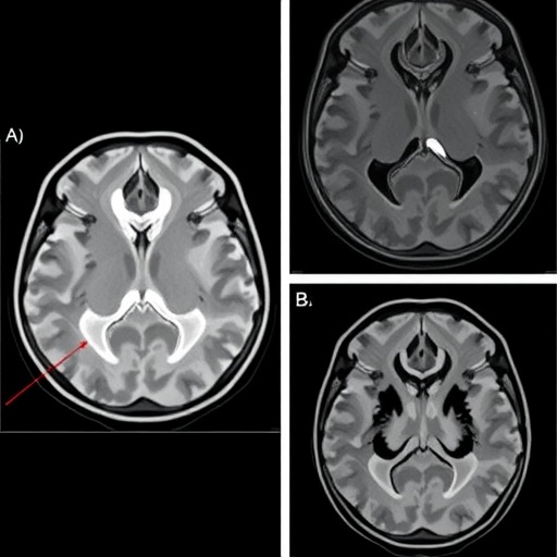
Image Credits: AI Generated
DOI: https://doi.org/10.1007/s00414-025-03686-w
Tags: advanced data mining techniquescartilage development stageschronological age determinationelbow MRI technologyethnic variability in age assessmentforensic age estimationforensic science advancementsjudicial processes and forensic evidencelegal age classification accuracymedical imaging in forensicsnon-invasive imaging methodsossification centers analysis