In the realm of chronic liver disease, a pressing global health challenge persists: the shortage of viable donor organs for transplantation and the limitations posed by patients who are too fragile for surgical interventions. Addressing this critical bottleneck, a team of pioneering engineers at the Massachusetts Institute of Technology (MIT) has unveiled a breakthrough technology—a novel approach to creating injectable “mini livers” that could revolutionize therapeutic options for liver failure patients. These engineered constructs aim to restore liver functions without the invasiveness and logistical constraints of traditional transplantation.
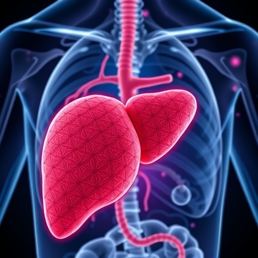
The liver, a powerhouse organ responsible for over 500 vital physiological functions, orchestrates complex tasks including detoxification, metabolic regulation, blood clotting, and immunological responses. Central to these roles are hepatocytes, the primary functional cells of the liver, which carry out most biochemical activities essential for life. Restoring or replacing the activities of these cells in end-stage liver disease has long been a therapeutic goal. However, conventional cell transplantation methods face significant hurdles—namely poor cell survival, inability to engraft effectively, and the need for invasive surgical implantation of supportive materials.
The team led by Professor Sangeeta Bhatia, an acclaimed figure in bioengineering and regenerative medicine, has addressed these challenges by innovating a unique microenvironment that supports hepatocyte survival, engraftment, and function post-transplantation. Their approach leverages cutting-edge microfluidic engineering to fabricate hydrogel microspheres—tiny, uniform spheres that encapsulate hepatocytes along with supportive fibroblast cells. These microspheres exhibit shear-thinning behavior, meaning they flow like a liquid under pressure but rapidly solidify once injected, creating a structured, cohesive network that mimics the extracellular matrix of liver tissue.
Functionally, these microspheres serve dual purposes. First, they provide a physical scaffold that maintains cell proximity and communication, crucial for restoring liver-specific functions. Second, they facilitate rapid vascularization by promoting blood vessel growth into the graft, a necessary feature for maintaining cell viability and enabling systemic delivery of hepatic proteins. The inclusion of fibroblast cells within this engineered niche further enhances vessel formation and hepatocyte support, as these cells secrete pro-angiogenic factors and create a nurturing environment for the transplanted cells.
Crucially, this technology eschews invasive surgery. The hydrogel-hepatocyte mixture is delivered via ultrasound-guided syringe injection into adipose tissue, such as the perigonadal fat pad in mice, allowing precise placement and minimization of patient trauma. Post-injection, the microspheres regain their solidity, stabilizing the newly formed graft and facilitating integration with host tissues. Ultrasound imaging also offers a non-invasive method to monitor graft survival and functionality longitudinally, advancing real-time assessment in clinical contexts.
In rigorous preclinical studies, the injected mini livers demonstrated remarkable stability and functionality for at least two months, maintaining viability and generating essential liver-specific proteins at levels comparable to native hepatocytes. Importantly, the neo-vascularized grafts displayed intimate blood vessel networks surrounding the transplanted cells, underscoring the success of the microenvironment in replicating physiologically relevant conditions. Such sustained function highlights the promise of this strategy as a potential long-term therapy that bolsters failing livers or serves as a supplementary “booster” organ, capable of sustaining patients while awaiting donor livers.
Beyond serving as a substitute for transplantation, this approach introduces a paradigm shift by creating “satellite livers” that can be deployed in multiple anatomical sites with adequate vascularization potential, such as near the spleen or kidneys. This flexibility not only broadens the applicability of hepatocyte grafts but also circumvents issues of local trauma and hepatic fibrosis often encountered in traditional transplantation sites. Furthermore, the injectable format allows for repeated administrations if necessary without the cumulative morbidity associated with multiple surgeries.
While immunosuppression remains a requisite barrier, the research team is actively exploring innovative strategies to circumvent immune rejection. These include engineering “stealth” hepatocytes that evade immune detection and utilizing the hydrogel microspheres as localized drug delivery systems for immunosuppressive agents—minimizing systemic side effects and enhancing the graft’s survival prospects. This thoughtful integration of immune modulation could elevate the clinical viability of the therapy significantly.
The implications of this research extend beyond liver diseases, as the conceptual framework of injectable, self-assembling cellular niches presents a prototype for regenerative therapies applicable to other organs and tissues. The successful harmonization of biomaterials engineering, cell biology, and medical imaging heralds a new era where minimally invasive, precision-guided cellular therapies can address organ failure more safely, efficiently, and accessibly.
Funding for this transformative research was provided by prominent institutions including the Koch Institute Support grant from the National Cancer Institute, the National Institutes of Health, and the Wellcome Leap HOPE Program. The confluence of interdisciplinary expertise and robust financial backing underscores the project’s potential to disrupt current therapeutic landscapes and improve outcomes for millions affected by liver disease worldwide.
In conclusion, MIT’s engineering of injectable mini livers marks a watershed moment in addressing the global challenge of liver failure. By harnessing the versatility of hydrogel microspheres and the biological functionality of hepatocytes, this novel bioengineered niche offers a less invasive, highly effective, and adaptable alternative to traditional transplantation. Continued advances in immune integration and clinical delivery promise to make satellite liver grafts a reality in the near future, profoundly impacting patient care and survival.
Subject of Research: Development of an injectable, image-guided hydrogel microsphere niche for hepatocyte transplantation to restore liver function.
Article Title: Image-Guided Injectable Niche for Hepatocyte Transplantation
News Publication Date: 3-Mar-2026
Web References:
10.1016/j.celbio.2026.100378
Image Credits: Bhatia Lab
Keywords
Biomedical engineering; Tissue engineering; Bioengineering; Liver; Hepatocytes; Regenerative medicine; Injectable therapy; Hydrogel microspheres; Cell transplantation; Organ regeneration
Tags: alternatives to liver transplantationbioengineered liver constructschronic liver disease treatment innovationshepatocyte cell therapy challengesinjectable liver tissue engineeringinjectable mini livers for liver failuremicroenvironment for hepatocyte survivalnon-surgical liver failure therapiesovercoming donor liver shortageProfessor Sangeeta Bhatia liver researchregenerative medicine for liver diseasesatellite liver technology MIT