
The human brain operates as a network of interconnected modules specialized for tasks that become less specialized as people age—a breakdown associated with memory loss and other forms of cognitive decline. Much remains unknown about the chemical and cellular machinery underlying this age-related brain decline, as well as ways in which genes, lifestyle, environment or medicine might alter its trajectory.
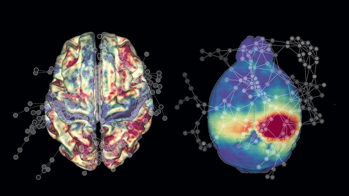
Now, scientists have uncovered commonalities in large-scale functional brain network decline between mice and humans, through scanning the brains of mice throughout their lifespans. Researchers used functional magnetic resonance imaging (fMRI) to scan the brains of 82 mice at several intervals from ages three to 20 months, roughly corresponding to ages 18 to 70 years in humans.
The findings suggest that the brains of humans and mice age in similar ways, information that may one day help scientists understand the factors underlying brain changes during aging and pinpoint mechanisms in humans that confer vulnerability to age-related brain decline, diseases, and disorders.
“By looking at mice, we can see if, say, a change in diet in their youth has an effect on them in old age, and we don’t have to wait 80 years for results as we would with humans,” said Itamar Kahn, PhD, principal investigator at Columbia’s Zuckerman Institute who is also an associate professor of neuroscience at Columbia’s Vagelos College of Physicians and Surgeons.
The team’s research was published in Proceedings of the National Academy of Sciences in the paper, “Correspondence of large-scale functional brain network decline across aging mice and humans.”
fMRI detects changes in blood flow to the brain. The researchers were able to capture images of the brains of mice while they are awake. The data suggest that aging mice, much like people, experienced a decline in how their different specialized brain modules interacted.
“The way the brain’s modules relate together as a whole is a measure of brain health that appears to apply similarly in both humans and mice,” said Ezra Winter-Nelson, a doctoral student in the lab of Gagan Wig, PhD, an associate professor of psychology at the University of Texas at Dallas.
The scientists also found significant differences between human and mouse brains. For instance, mouse brain modules communicated less with each other than human ones.
“We think the greater integration that humans have across their brain networks may contribute to aspects of cognition that are especially developed in humans,” Wig said.
In addition, the human decline in brain module specialization was faster than in mice. “So while we as humans have this ability to integrate information across more widely distributed parts of the brain, that may leave us more vulnerable to brain and cognitive decline when compared to mice,” Wig added.
The researchers noted that one limitation to the study is that they investigated only one type of lab mice. “We know there are other types of mice that show variability in how they respond to aging,” Kahn said. “So we want to look at other types of mice to understand how genetics affect trajectories of aging.”
Previous mouse neuroscience research has drawn criticism for often not possessing clinical relevance in humans. Much of that prior work looked at changes seen at the cellular level. “What we’re doing is looking at the brain at the network level,” Kahn noted. “We believe that looking at both the cellular and network level in mice may prove better for developing therapeutic approaches that actually work in humans.”