For decades, microtubules, slender dynamic filaments integral to a cell’s cytoskeleton, were regarded predominantly as passive structural elements, serving as mere mechanical scaffolds that maintain cellular shape and provide tracks for intracellular transport. However, a groundbreaking study published in Science Advances has radically transformed this perspective, uncovering a vital, previously unappreciated signaling capacity of microtubules. This research reveals that microtubules actively regulate enzymatic activity by modulating the spatial configuration of substrate proteins, thereby orchestrating crucial events during cell division with exquisite precision.
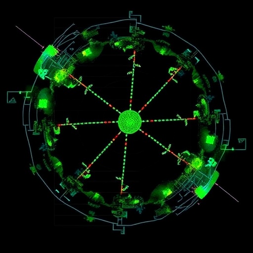
The process of faithful chromosome segregation during mitosis has long fascinated cell biologists. Chromosomes must be accurately duplicated and then equally distributed into two daughter cells, a feat achieved through the mitotic spindle, a complex assembly predominantly composed of microtubules. The attachment of microtubules to chromosomes occurs at specialized structures called kinetochores, located at the centromeres of chromosomes. These microtubule-kinetochore attachments are critical for maneuvering chromosomes to opposite poles of the dividing cell. Yet, the inherent stochastic nature of these attachments results in frequent errors. Erroneous attachments can lead to chromosomal instability, aneuploidy, and are strongly implicated in oncogenesis.
Central to the surveillance and correction of these attachment errors is the multifunctional enzyme Aurora B kinase. This kinase delicately balances the destabilization of incorrect kinetochore-microtubule connections while maintaining the integrity of the spindle apparatus. Aurora B selectively phosphorylates kinetochore substrates to weaken improper attachments, enabling their release and corrective reattachment, but simultaneously inhibits MCAK, a microtubule depolymerase that would otherwise destabilize the entire spindle structure. Deciphering how Aurora B distinguishes correct from incorrect attachments has long posed a challenge for molecular cell biology.
Earlier mechanistic models posited that correct amphitelic bi-oriented attachments physically pull the kinetochore away from the centromeric concentration of Aurora B, attenuating its substrate phosphorylation. However, accumulating evidence demonstrated the presence of active Aurora B at kinetochores and its ability to bind microtubules directly, suggesting a more nuanced mechanism beyond mere spatial separation. Hypothesizing that microtubules themselves modulate Aurora B’s access to its targets, the team led by Hironori Funabiki advanced the concept that microtubule binding protein geometries influence kinase activity.
To empirically test this hypothesis, the researchers performed elegant in vitro reconstitution using purified components, including Aurora B kinase, the regulatory Chromosomal Passenger Complex, the kinetochore-associated Ndc80 complex, and MCAK. The Ndc80 complex serves as a pivotal microtubule attachment site within the kinetochore, anchoring chromosomes to the spindle. Strikingly, when Ndc80 was pre-bound to microtubules, Aurora B displayed diminished ability to phosphorylate it, implying that the microtubule scaffold obstructed access. Conversely, MCAK remained readily phosphorylatable even when bound to microtubules, highlighting differential accessibility influenced by substrate microtubule interactions.
Employing cryo-electron microscopy offered unprecedented molecular insight into this phenomenon. The team discerned that Ndc80 molecules, upon microtubule binding, cluster densely along the filament in discrete arrays. This oligomerization effectively conceals phosphorylation sites from Aurora B, serving as a physical barrier that impedes kinase engagement. MCAK, by contrast, associates with microtubules without such clustering, leaving phosphorylation sites exposed and susceptible to enzymatic modification. This distinct spatial arrangement functions as a molecular switch—mediated by microtubules—that dictates substrate susceptibility to Aurora B.
This mechanism elegantly resolves the paradox of how Aurora B can both target and spare proteins during mitosis. When an Ndc80 complex is not yet clustered, Aurora B phosphorylates it, weakening potential erroneous attachments. Once correct microtubule attachments mature into clustered Ndc80 arrays, Aurora B is physically precluded from further phosphorylation, thereby stabilizing the connection and protecting spindle integrity. MCAK remains a consistent target, permitting Aurora B to inhibit microtubule depolymerization selectively and maintain spindle architecture.
The functional importance of Ndc80 clustering was further substantiated in living cells engineered to express mutant Ndc80 incapable of clustering despite normal microtubule binding. These mutants exhibited compromised kinetochore-microtubule stability, leading to improper chromosome segregation and cell division errors, underscoring the physiological relevance of this mechanochemical regulatory system.
Collectively, these findings redefine the role of microtubules from passive architectural filaments to dynamic regulators that spatially dictate enzymatic activities critical for mitosis. By sculpting substrate geometries, microtubules effectively control the temporal and spatial dynamics of Aurora B kinase activity, ensuring fidelity in chromosome segregation. This sophisticated quality-control mechanism exemplifies the integration of mechanical and biochemical signals within the cell to prevent chromosomal instability.
Implications of this research extend into oncology, where chromosomal missegregation is a hallmark of tumorigenesis. A thorough molecular understanding of the Ndc80 clustering and Aurora B regulation pathway opens avenues to explore why this quality control system fails in cancer cells, potentially guiding therapeutic interventions targeting mitotic regulation.
As Yiming Niu, a key researcher in the study, reflects, “Microtubules are no longer mere cellular scaffolds; they are active directors of biochemical processes essential for life.” This breakthrough not only enriches the fundamental knowledge of mitotic regulation but may also inspire future investigations into cytoskeletal regulation of enzyme-substrate interactions across diverse cellular contexts.
The combination of cutting-edge biochemical reconstitution, high-resolution structural analysis, and functional cell biology in this study exemplifies how multidisciplinary approaches can unravel the intricate control mechanisms underpinning cell division, a cornerstone of life itself.
Subject of Research: Regulation of enzymatic activity by microtubule protein geometry during mitosis
Article Title: Microtubules guide Aurora B substrate geometries for accurate chromosome segregation
News Publication Date: 25-Mar-2026
Web References: DOI: 10.1126/sciadv.aea2112
Image Credits: Laboratory of Chromosome and Cell Biology at The Rockefeller University
Keywords: Microtubules, Cell division, Aurora B kinase, Ndc80 complex, Chromosome segregation, Kinetochore, Mitotic spindle, MCAK, Cytoskeleton, Enzyme regulation, Cryo-electron microscopy, Chromosomal instability
Tags: active regulation of enzymatic activity by microtubulesAurora B kinase role in mitosischromosomal stability and mitosischromosome segregation during mitosiscorrection of chromosome attachment errorsdynamic cytoskeleton in chromosome distributionmechanisms of aneuploidy preventionmicrotubule signaling in cell divisionmicrotubule-kinetochore attachmentsmicrotubules and oncogenesis linkmicrotubules in mitotic checkpointmitotic spindle function and structure