Sponsored content brought to you by
![]()
At the 2026 Advances in Genome Biology and Technology (AGBT) meeting in Orlando, Bruker Spatial Biology unveiled the Cell-Scape™ XR platform, its next-generation spatial proteomics system. As part of Bruker Spatial Biology’s approach to capturing the full complexity of biology, the debut signals something larger than a routine product launch. In a comprehensive ecosystem of spatial biology tools designed to span DNA, RNA, and protein, multiplexed immunofluorescence (mIF) is not simply another modality—it is the benchmark that anchors and validates all other spatial measurements.
Over the past decade, high-plex single cell and spatial transcriptomics have transformed biological discovery. Platforms such as CosMx® Spatial Molecular Imager have enabled single-cell and subcellular mapping of the whole transcriptome. Yet even as RNA-based tools grow in power and scale, proteins remain the functional currency of the cell—and the layer most directly tied to phenotype and clinical decision making. Perhaps more importantly, mIF serves as a natural extension to clinical immunohistochemistry (IHC), a foundational modality in clinical tissue biology for more than a century.
Traditional mIF workflows have often been hindered by the following limitations: harsh stripping or signal removal protocols that can damage tissue morphology and compromise epitope integrity; limited antibody compatibility that constrains panel design and customization; and slow throughput that prevents study of large clinical cohorts. Bruker Spatial Biology engineered the CellScape XR platform to address these constraints.
The CellScape XR system is designed to deliver highly quantitative spatial proteomics. By combining advances across the instrument, assay and workflow, the platform enables rapid, robust and flexible mIF without compromising tissue integrity. At its core is Bruker’s EpicIF™ technology, which enables rapid whole-slide signal removal between staining cycles without harsh antibody stripping. This approach preserves epitope integrity, expands antibody compatibility and supports flexible panel design through VistaPlex™ Multiplex Assay Kits and the new VistaFlex™ universal antibody labeling. The result is true quantitative spatial phenotyping with greater assay flexibility and more than twice the throughput of prior CellScape platforms.
Researchers working at the intersection of RNA and protein spatial biology underscore the importance of this approach.
Parambir Dulai, MD, associate professor of medicine at Northwestern University, whose lab integrates spatial transcriptomics with multiplexed protein analysis, emphasizes the complementary nature of the approaches. “Spatial transcriptomics gives us extraordinary breadth in understanding tissue states,” Dulai notes. “But CellScape gives us the ground truth at the protein level. It’s essential for validating cell phenotypes and ensuring that our RNA-based findings reflect functional biology.”
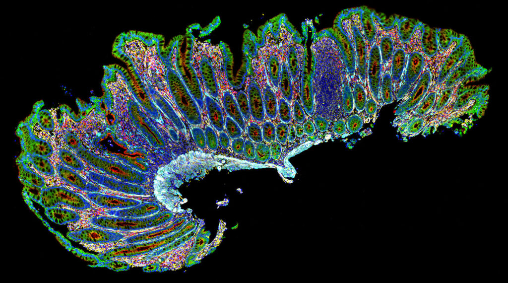
Dulai highlights an example in his study of gastrointestinal disease, where the delineation of T cell subsets based on FoxP3 expression is important. “With high-fidelity imaging, we can distinguish low-, medium-, and high-expressing FoxP3 T cell populations within the same tissue,” he says. “That level of quantitation enables us to better define regulatory T cell states, their spatial relationships, and the role they play in Crohn’s disease and ulcerative colitis.”
The CellScape XR platform’s improvements in signal consistency, throughput, and customization aim squarely at the demands of translational research. By expanding antibody compatibility, doubling throughput, and avoiding harsh assay chemistry, the CellScape XR system aligns mIF with the scale and rigor demanded by modern translational and clinical research.
As the field of spatial biology matures, researchers are generating increasingly complex multiomic datasets. Yet as complexity increases, so does the need for biological grounding. Protein expression remains the decisive layer of biology for defining cellular phenotype and functional state. In that context, mIF is not a secondary validation step—it is the reference standard.
With the CellScape XR system, Bruker reinforces its broader portfolio strategy: Best-in-class, high fidelity solutions across the full complexity of biology. By delivering flexible subcellular proteomics for rapid and quantitative spatial phenotyping—within an ecosystem that includes visualization of the 3D genome and complete spatial transcriptomics—CellScape XR provides the protein information required to translate spatial biology from discovery into clinical impact.

Learn more www.brukerspatialbiology.com.