In a groundbreaking advancement set to revolutionize biomolecular imaging, a team of researchers led by Shen, B., Zeng, Z., and Li, H. has unveiled an innovative microscopy technique that performs simultaneous dual-Raman-shift scanning-free stimulated Raman scattering (SRS) imaging. Published in the prestigious journal Nature Communications in 2026, this study represents a pivotal leap toward label-free, high-speed biochemical visualization at the cellular and molecular levels. The novel method addresses longstanding challenges in Raman microscopy, delivering unprecedented imaging speed and molecular specificity without relying on fluorescent or other external labels, which often perturb biological samples.
Stimulated Raman scattering microscopy has emerged as a powerful tool for visualizing molecular vibrations intrinsic to biomolecules, enabling researchers to directly observe biochemical compositions in live cells and tissues. However, traditional SRS approaches rely heavily on sequential scanning, which limits temporal resolution and hampers real-time observations of dynamic biological processes. Moreover, extracting multiple Raman spectral signatures typically necessitates serial acquisition steps, prolonging imaging sessions and increasing photodamage risks. The present research circumvents these bottlenecks by pioneering a simultaneous acquisition strategy. This dual-Raman-shift technique captures multiple vibrational frequencies concurrently, enabling more comprehensive biochemical mapping rapidly and non-invasively.
At the core of the innovation lies a sophisticated optical configuration that generates two synchronized stimulated Raman signals at distinct Raman shifts without requiring mechanical scanning. The team harnessed a tailored laser modulation scheme combined with an advanced detection framework, allowing the selective excitation and detection of two molecular vibrational modes simultaneously. This approach dramatically accelerates the imaging process, providing real-time, multiplexed insights into biomolecular structures and concentrations. The absence of scanning hardware not only simplifies the instrumentation but also enhances system robustness and stability, vital for sensitive biological assays.
One of the most striking features is the method’s ability to differentiate between chemically similar molecules within complex cellular environments based on their distinct vibrational fingerprints. By capturing dual Raman signals simultaneously, the system elegantly deciphers subtle biochemical differences—such as between lipid and protein domains—allowing researchers to visualize cellular metabolism, structural organization, and pathological transformations with microscopic precision. Importantly, this label-free technique maintains native biomolecular states, avoiding artifacts induced by fluorescent markers or dyes and enabling long-term live-cell imaging.
The implications for biomedical research are profound. In fields ranging from cancer biology to neuroscience, rapid, detailed molecular imaging has been hampered by technical constraints. This dual-Raman-shift SRS microscopy bridges that gap, empowering scientists to observe cellular biochemistry dynamics in vivo with previously unattainable speed and accuracy. The scanning-free design is particularly advantageous for studying fast biological events—such as neurotransmitter release, metabolic fluxes, or membrane remodeling—facilitating discoveries into fundamental cellular mechanisms and disease pathways.
Technically, the researchers implemented a pulse shaping and modulation technique to generate synchronized pump and Stokes beams tailored to excite vibrational modes corresponding to distinct biomolecular bonds. By integrating a lock-in detection scheme capable of demodulating the dual-frequency signals, the setup achieved high sensitivity and specificity. The careful orchestration of laser parameters ensures minimal photothermal damage, preserving cellular viability even during extended imaging sessions. This makes it feasible to monitor live biological specimens longitudinally, providing dynamic biochemical snapshots with exquisite spatial and temporal resolution.
Furthermore, the system accommodates integration with conventional microscopes and can be adapted for in vivo imaging applications. This versatility opens doors to translational research, where rapid, label-free molecular imaging could enhance diagnostics and therapeutic monitoring. For example, in oncology, delineating tumor margins and metabolic heterogeneity in real-time could inform precision surgery or drug delivery strategies. In neuroscience, mapping neurotransmitter distributions across brain regions with millisecond temporal resolution could unravel complex signaling networks.
Another compelling aspect is the dual Raman shift strategy’s impact on data throughput and analysis. Simultaneous acquisition reduces the volume of raw images needed to capture comprehensive molecular information, easing computational burdens and expediting data interpretation. Coupled with advanced machine learning algorithms, this approach promises rapid biomolecular classification and anomaly detection, facilitating automated, high-content analysis pipelines indispensable for large-scale biological studies.
The team’s findings also underscore the broader potential of multi-frequency, scanning-free stimulated Raman technologies to extend beyond just dual Raman shifts. Future developments might incorporate multiplexed configurations capturing even richer vibrational spectra concurrently, further enhancing multiplexing capabilities without compromising resolution or acquisition speed. Such advancements could redefine conventional paradigms in vibrational imaging, establishing a new class of microscopy techniques tailored for complex, dynamic biological systems.
From an engineering perspective, the removal of mechanical scanning components mitigates common issues related to system wear, vibration-induced artifacts, and alignment drift, thereby ensuring long-term measurement reproducibility—a critical factor in both research and clinical environments. The compact and streamlined design enables miniaturization potentials, possibly paving the way for portable or handheld Raman imaging devices that bring cutting-edge molecular imaging directly to bedside or field settings.
In summary, the simultaneous dual-Raman-shift scanning-free stimulated Raman scattering microscopy introduced by Shen and colleagues opens new horizons in label-free biomolecular imaging by uniting speed, sensitivity, and multiplexed chemical specificity into a single, robust platform. This technique propels vibrational microscopy into real-time, dynamic biological exploration mode, poised to catalyze breakthroughs across diverse science and medical disciplines. The combination of technical ingenuity and profound applicability marks this development as a seminal milestone in optical bioimaging evolution.
Continued research inspired by this work will likely delve into expanding Raman frequency coverage, refining laser modulation schemes, and optimizing detection electronics to further boost performance. Moreover, interdisciplinary collaborations integrating this imaging modality with other analytical techniques, such as mass spectrometry or electron microscopy, could enrich multimodal datasets to capture biomolecular landscapes with unparalleled depth. The ongoing evolution of scanning-free SRS microscopy promises a future where live, comprehensive biochemical mapping is routine, transforming our understanding of life’s molecular machinery in both health and disease.
This transformative approach to label-free biochemical imaging owes its success to meticulous engineering, innovative photonics design, and deep understanding of molecular vibrational physics. By circumventing the limitations of scanning mechanisms and sequential spectral acquisition, the simultaneous dual-Raman-shift SRS microscopy stands as a beacon of next-generation vibrational imaging technology, enabling researchers to visualize the untold stories encoded within biomolecular vibrations with clarity and immediacy never before realized.
Subject of Research: Simultaneous dual-Raman-shift scanning-free stimulated Raman scattering microscopy for label-free biomolecular imaging
Article Title: Simultaneous dual-Raman-shift scanning-free stimulated Raman scattering microscopy for label-free biomolecular imaging
Article References:
Shen, B., Zeng, Z., Li, H. et al. Simultaneous dual-Raman-shift scanning-free stimulated Raman scattering microscopy for label-free biomolecular imaging. Nat Commun (2026). https://doi.org/10.1038/s41467-026-73391-8
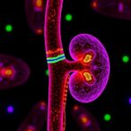
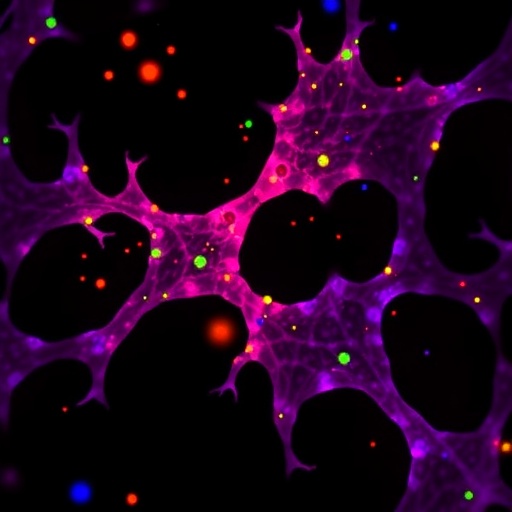
Image Credits: AI Generated
Tags: advanced stimulated Raman scattering methodsdual-Raman-shift microscopyhigh-speed biochemical visualizationlabel-free biomolecular imaginglive cell biochemical mappingmolecular specificity in microscopymultiplexed Raman spectral acquisitionnon-invasive cellular imaging techniquesoptical configuration for Raman microscopyreal-time biomolecular dynamicsscanning-free stimulated Raman scatteringsimultaneous dual-frequency Raman imaging