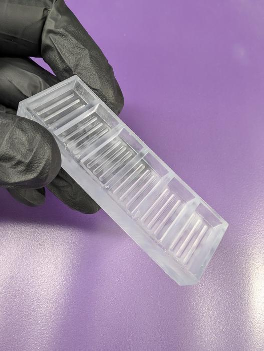
![Special grooved trays developed at Cincinnati Children's were a key part of a novel process to produce larger, faster-growing gut organoids for use in medical research and potential human tissue repair. [Cincinnati Children's]](https://www.genengnews.com/wp-content/uploads/2026/05/Low-Res_molds.jpg "Low-Res_molds")
Researchers at Cincinnati Children’s Hospital Medical Center and Nantes Université in France have designed 3D-printed scaffolding trays that will reportedly allow scientists to produce larger versions of functional human gut organoids twice as fast as previous methods—and these organoids grow their own nerve cells.
This improved technology could help accelerate production of human mini-organ tissues that are large enough to be useful in patching damage or restoring diminished functions of a person’s small intestine, stomach, or colon. Such tissues also would be valuable for future disease studies and to more accurately evaluate organ damage risks linked to oral medications, according to the investigators.
Details of the study “Large-scale and innervated functional human gut tissues for transplantation via transient spheroid confinement” appear in Nature Biomedical Engineering.
Using a confined culture system (CCS), the team grew small intestine, colon, and stomach organoids from tiny spherical forms into centimeter-scale tubular forms nearly 10 times larger than previous methods. Also, unlike methods that require a complex effort to introduce nerve cells, these organoids develop a nervous system on their own.
“By reaching transplantation maturity twice as fast and developing their own functional nerves, these organoids demonstrate how engineering principles can drive biological innovation,” said staff investigator Holly Poling, PhD. “Our confined culture system is more than a production method; it’s a scalable, flexible platform for building complex human tissues.”
New production system prompts faster growth
Experts at Cincinnati Children’s Center for Stem Cell & Organoid Medicine (CuSTOM) have been making miniature versions of digestive system organs for more than 15 years, working on improving the sophistication of the lab-grown tissues. More recently, the team has been developing methods to make enough customized tissue to transplant into patients to help patch organ damage or restore diminished specialized functions.
The new technique uses 3D printing technology to make tray-like scaffolding molds from surgical resin, then filling the molds with degassed polydimethylsiloxane—a flexible rubber-like type of silicone.
The new trays contain grooves designed to confine a collection of sphere-shaped organoids into a row, which encourages the spheroids to fuse together and mature. The fusions occur within a special mix of nutrients and other ingredients that support initial growth from induced pluripotent stem cells (iPSCs) into more complex organoids.
By day six, the discrete spheroids develop into unified constructs along the grooves of the trays. These are moved into another hydrogel medium for continued growth for another eight days.
By day 14, the organoid constructs have produced all the cell types and structures that previously required 28 days to achieve. These tissues are then transplanted into rodents that are genetically modified to minimize rejection risk.
All of the transplanted tissues engrafted in rodents, the co-authors state. After growing in the rodents, the team produced as much as eight cm of functioning small intestine tissue, compared to approximately one cm of tissue using previous protocols. Not only were the structures much larger than previous methods, but now their neuromuscular function was also similar to native human tissue, representing a major advance.
“We are now able not only to generate complex gastrointestinal organoids at scale, but also to guide their differentiation into functional tissues with integrated enteric neuronal networks,” noted senior author Maxime Mahe, PhD. “By leveraging a defined growth environment, the intrinsic self-organization capacity of the cells drives the formation of tissue structures that closely resemble the human gastrointestinal tract.”
Jim Wells, PhD, a study co-author and chief scientific director at CuSTOM says the new technology overcomes key barriers to scale and function in organoid research and biomanufacturing.
“This platform’s simplicity, reproducibility, and versatility make it accessible for widespread adoption,” said Wells. “In addition, the emergence of a self-organized nervous system within these organoids is particularly important for further studies of neurodevelopmental disorders.”
Another step closer to human clinical trials
Michael Helmrath, MD, a surgeon-scientist at Cincinnati Children’s who co-directs CuSTOM, has been working for more than a decade to develop intestine organoids sophisticated enough for transplantation in human patients.
In 2017, Helmrath and colleagues demonstrated how to combine neural crest cells with intestinal tissue cells in a layered process to make the first human organoids with nerve function. His team also showed how intestine organoids could be grown larger by implanting them in a mouse to provide a blood supply. Ever since, intestine organoids have been getting more sophisticated, including versions with immune cells in addition to the specialized organ cells and nerves.
Now the new process—involving rats instead of mice—produces larger amounts of tissue.
“It is still not possible to grow complete, full-sized human organs in some sort of tank, but research like this has produced significant amounts of tissue that can be matched directly to individual patients,” explains Helmrath. “We believe such tissues, once transplanted, would further grow and multiply as part of the patient’s own organ to restore functions.”
More research and development is needed before “CCS organoids” will be ready for human clinical trials, according to Helmrath. But if successes continue, organoid medicine may allow more infants and children with dysfunctional organs to be treated without ever needing a full organ transplant.