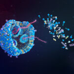
Researchers showed how what appears to be a tangle of DNA is actually organized into a structure that coordinates thousands of genes to form a sperm cell. The work, published as two papers in Nature Structural and Molecular Biology, could improve treatment for fertility problems and developmental disorders.
“We are finding the 3D structure of the genome,” said Satoshi Namekawa, PhD, professor of microbiology and molecular genetics at the University of California, Davis, and senior author of one of the papers: “CTCF-mediated 3D chromatin sets up the gene expression program in the male germline.”
“This is really showing us how the genomic architecture guides development.”
Genes can be physically close to the enhancer DNA switches that turn them on and off even if they are far apart in the DNA sequence. To understand how genes are turned on and off to make different cell types, it is essential to figure out how DNA is folded, and which genes and enhancers are paired together.
Memories of cells
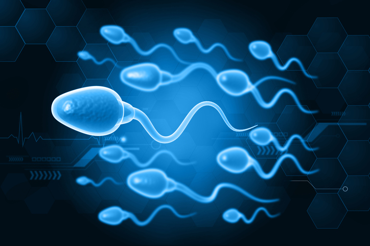
“In a mouse or human embryo, the cells that will one day produce sperm or eggs are already earmarked for that future purpose,” explained Namekawa. These primordial germ cells are initially bipotent (able to become either sperm or eggs). But while the embryo is still in the womb, its bipotent cells commit to one path or another and once they cross that threshold, they cannot go back.
“Cells have a sort of memory,” continued Namekawa. “But we don’t know how that memory works. We are trying to understand how this male fate is acquired.”
Namekawa and his students used the “Hi-C” approach to identify the proximity junctions where far-flung parts of the genome are paired up next to one another. Using a computer to analyze those paired locations, the team was able to see how the entire string of DNA is looped and folded.

In the new studies, Yuka Kitamura, PhD, UC Davis postdoctoral fellow, identified two proteins in germ cells that establish this cellular memory. The first, called SCML2, disconnects junctions and allows the DNA to unfold and loosen, preparing it to be reorganized in the next stage of sperm cell development. Another protein, CTCF, attaches to locations of super-enhancer DNA and pairs them up with genes that they will later turn on during sperm development. This sets up a new structure that cements the cell’s future fate as a sperm cell.
The companion paper, “ZBTB16/PLZF regulates juvenile spermatogonial stem cell development through an extensive transcription factor poising network,” demonstrates that well before the germ cells enter meiosis, CTCF and other proteins bookmark thousands of locations in the genome. This paper was led by postdoctoral fellow Chongil Yi, PhD, and Bradley Cairns, PhD, professor and chair of oncological sciences at the University of Utah School of Medicine and an investigator at the Howard Hughes Medical Institute. It was published in collaboration with Namekawa and Kitamura.
These discoveries could have important medical implications, including diagnostic tests for causes of infertility linked to genome folding, according to the scientists, who add that the discoveries could also help scientists working on stem cell therapies, since coaxing a stem cell to become a neuron or heart cell requires shifting it from one genetic program to another—each defined by a particular 3D genome structure.
“We are uncovering the language of cell memory and cell fate,” Namekawa said.