In a groundbreaking study recently published in PhotoniX Life, researchers have unveiled a revolutionary label-free technique that offers an unprecedented window into the intricate world of subcellular dynamics. By harnessing the capabilities of wide-field interferometric scattering (iSCAT) microscopy paired with power spectral density (PSD) analysis, this novel method delivers an advanced approach for mapping and visualizing the nuanced motions occurring within living cells. The implications of this study reach far beyond traditional cell imaging, promising transformative impacts across mechanobiology, cancer diagnostics, and stem cell research.
Interferometric scattering microscopy has long held promise as a label-free optical technique capable of detecting nano-scale cellular events without the need for fluorescent dyes or contrast agents. Capitalizing on the interference between scattered light from the sample and a reference beam, iSCAT achieves extraordinary sensitivity to minuscule refractive index variations inside cells. However, prior implementations predominantly focused on static imaging or limited dynamic analyses. The present work transcends these constraints by integrating PSD analysis, which examines temporal fluctuations in the iSCAT signal, thereby capturing the dynamic landscape of intracellular motion over a broad frequency range.
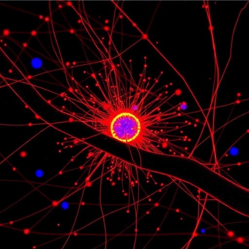
At the core of this study lies the detailed interrogation of pixel-wise PSDs extracted from wide-field iSCAT images. By fitting these PSDs to an inverse-power-law model in the bandwidth from 30 Hz to 1,250 Hz, the researchers derived what they call spectral exponent maps. These maps provide a spatially resolved depiction of the strength and nature of subcellular dynamics. Essentially, these spectral exponents quantify how fluctuations vary with frequency across the cell, offering an insightful metric that correlates directly with underlying biological processes like cytoskeleton remodeling, organelle transport, and molecular trafficking.
The power-law behavior detected in the PSDs is particularly noteworthy because it mirrors complex physical phenomena known to characterize non-equilibrium biological systems. Subcellular components do not move randomly but exhibit correlated motions influenced by metabolic energy, biochemical reactions, and mechanical forces. The spectral exponent thus encapsulates these rich mechanobiological signatures in a succinct, quantitative manner—a feat that conventional microscopy techniques struggle to achieve without invasive labeling or genetic modification.
Using this approach, the researchers successfully distinguished between cells in different physiological states. For instance, mitotic cells—those actively dividing—displayed distinct spectral exponent signatures compared to cells in interphase, the quiescent phase of the cell cycle. This discrimination reflects the heightened cytoskeletal rearrangements, mitotic spindle formation, and dynamic organelle positioning characteristic of mitosis. Such ability to detect cell cycle phase purely from intrinsic optical signals represents a leap forward for label-free cell biology studies.
Moreover, the method enabled the differentiation of live cells from those undergoing apoptosis, or programmed cell death. Apoptotic cells exhibited altered spectral exponent profiles, indicative of dramatic shifts in intracellular dynamics such as membrane blebbing, cytoskeletal disassembly, and organelle fragmentation. The capacity to optically identify apoptosis without fluorescent markers provides a powerful tool for real-time monitoring of cell viability in pharmacological assays and cancer research.
Perhaps most striking is the technique’s application in oncology. When applied to thyroid cancer cells, the researchers identified spectral exponent variations corresponding to malignancy grade. Cells originating from more aggressive tumor subtypes showed markedly different dynamic signatures compared to benign or less malignant cells. This suggests that the intrinsic optical fluctuations captured by iSCAT and PSD analysis may serve as a novel, non-invasive biomarker for cancer diagnosis, prognosis, and therapeutic response monitoring.
This advancement also holds promise for stem cell biology. Stem cells exhibit distinctive mechanical properties and dynamic behaviors during differentiation and self-renewal. By mapping these characteristics label-free, the technique offers an innovative means to assess cell potency and developmental state. Such capability could accelerate stem cell-based regenerative medicine by providing rapid, quantitative feedback on cell quality without perturbing native physiology.
Technically, the wide-field iSCAT system utilized offers rapid imaging speeds and mesoscopic field of view, enabling simultaneous capture of thousands of pixels and thereby facilitating robust statistical analysis of PSDs. The inverse power-law fitting procedure not only condenses complex temporal fluctuations into single scalar values per pixel but also allows high-resolution spatial mapping across whole cells, generating intricate heatmaps reflecting localized dynamic hotspots.
The implications extend beyond biology. Understanding mechanobiology within cells at this level could inform novel biomaterial designs and contribute to synthetic biology efforts aimed at designing artificial cellular components. The ability to monitor intracellular dynamics purely through scattered light interference also opens avenues for label-free diagnostics in clinical settings, potentially reducing reliance on exogenous contrast agents that can influence cell behavior or elicit toxicity.
Crucially, the methodology is entirely label-free and non-destructive, preserving cell viability and avoiding phototoxic effects often associated with fluorescent microscopy. This is paramount for live-cell studies over extended timescales, longitudinal monitoring in drug development pipelines, and sensitive clinical samples. The broad frequency range analyzed captures diverse motion scales—from organelle-level nanometer oscillations to cytoskeletal rearrangements—offering a holistic portrait of subcellular activity.
While the technique currently focuses on cultured cell models, future extensions could enable in vivo investigations or three-dimensional iSCAT imaging with tomographic reconstructions. Integration with machine learning algorithms may further enhance classification accuracy of cell states and disease subtypes from spectral exponent datasets, setting the stage for automated diagnostics and high-throughput screening.
In summary, this pioneering study presents a powerful optical instrumentation and data analysis framework that transforms subcellular dynamics into quantifiable maps revealing biological state, disease progression, and mechanistic insights without the need for artificial labels. By exploiting the rich temporal complexity of interferometric scattering signals, it bridges physics and cell biology, offering a versatile tool poised to ignite new frontiers in life sciences and medical research.
Subject of Research: Subcellular dynamics and label-free cellular imaging using wide-field interferometric scattering microscopy and power spectral density analysis.
Article Title: Not provided.
News Publication Date: Not provided.
Web References: Not provided.
References: Not provided.
Image Credits: Not provided.
Keywords: interferometric scattering microscopy; iSCAT; power spectral density; PSD analysis; subcellular dynamics; label-free imaging; spectral exponent maps; mechanobiology; cancer diagnostics; thyroid cancer; apoptosis; mitosis; stem cell assessment.
Tags: cancer cell diagnostics microscopydynamic cellular behavior analysisintracellular motion mapping techniqueslabel-free interferometric scattering microscopymechanobiology imaging advancementsnano-scale cellular fluctuations detectionnon-fluorescent cellular imaging methodspower spectral density analysis in cellsrefractive index variations in cellsstem cell research imaging toolssubcellular dynamics visualizationwide-field iSCAT imaging