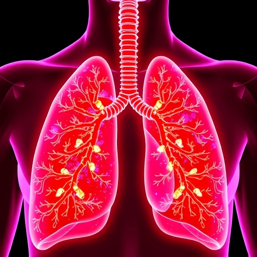
A groundbreaking study from NYU Langone Health and its Perlmutter Cancer Center elucidates a novel mechanism by which lung tumors induce cachexia—a debilitating wasting syndrome marked by profound weight loss and muscle depletion in cancer patients. Despite its prevalence and detrimental impact on treatment eligibility and survival, cachexia’s underlying biology has remained poorly understood, hindering effective therapeutic development.
The research, published in Science, focused on genetically engineered mouse models mimicking common human lung cancer subtypes. Intriguingly, only mice harboring tumors lacking the tumor suppressor gene LKB1 developed severe cachexia, characterized by loss of both fat and muscle mass driven by diminished appetite. This cachectic phenotype was not correlated with larger or more aggressive tumors, hinting at an alternative pathological driver.
A striking discovery emerged when these LKB1-deficient mice were fed a high-fat, high-calorie diet—a common clinical recommendation to combat weight loss. Rather than improving their condition, the dietary switch exacerbated weight loss, suppressed appetite further, and accelerated mortality. Analysis of lung tumor microenvironments revealed that this worsening was linked to elevated prostaglandin E2 (PGE2), a lipid mediator known to potentiate inflammation.
Remarkably, PGE2 was found confined within the lung tissue, with no corresponding rise in systemic circulation, suggesting a localized paracrine effect. Building on recent findings from pulmonary infection models, the researchers posited that tumor-produced PGE2 acts on sensory neurons in the lung to relay anorexic signals to the brain via the vagus nerve. Experimental interruption of vagal nerve signaling—either surgically or genetically—reversed appetite suppression and mitigated cachexia symptoms in affected mice.
Pharmacological and dietary interventions targeting PGE2 production, including administration of NSAIDs and omega-3 rich fish oil, resulted in improved survival outcomes and attenuated weight loss, even under the high-fat dietary regimen. These interventions did not impact tumor growth, highlighting a distinct cachexia pathway separable from tumor proliferation.
Corroborating the murine data, lung fluid samples from human lung cancer patients demonstrated elevated PGE2 concentrations in individuals suffering from cachexia, underscoring the potential translational relevance of targeting this pathway. This work suggests that sensory neuron-mediated communication within the tumor microenvironment is a critical driver of cancer-associated cachexia.
This paradigm-shifting research reveals that altering neural signaling pathways, rather than solely focusing on systemic inflammation or tumor control, could offer new avenues for preserving patient strength and improving tolerance to cancer therapies. It opens the door for novel multidisciplinary strategies that incorporate neuronal, metabolic, and inflammatory processes in combating cachexia—an urgent unmet need in oncology care.
Further investigations by this international consortium, Cancer Grand Challenges, aim to deepen understanding of neuronal signaling roles in cancer syndromes and to identify targeted interventions to alleviate cancer-associated suffering.
Subject of Research: Animals
Article Title: A dietary switch promotes sensory neuron–dependent cancer-associated cachexia
News Publication Date: July 2, 2024
Web References: 10.1126/science.adz4196
Keywords: Lung cancer, Cachexia, Prostaglandin E2, Vagus nerve, Sensory neurons, Inflammation, LKB1, Cancer-associated weight loss
Tags: cachexia mechanismcancer cachexia treatment challengesinflammation and cancer progressionLKB1 gene deficiencylocalized lipid mediators in tumor biologylung cancerlung tumor-induced appetite suppressionmuscle and fat depletion in cancernervous system hijackingprostaglandin E2 in cancertumor microenvironmenttumor-induced weight loss