Improving the cultivation of large bioprinted tissues could help make laboratory-grown tissue models more reliable for drug development and regenerative medicine. At the International Society for Cell & Gene Therapy (ISCT) 2026 annual meeting in Dublin, Elliot Cowles, a doctoral student in bioprocess engineering at Université Claude Bernard Lyon, and colleagues presented a perfusion-based culture system, which was described in Cytotherapy, that addresses this challenge. Although biofabricated tissues are increasingly used for in vitro research, many remain relatively small and are typically grown under static conditions that limit scalability and consistency.
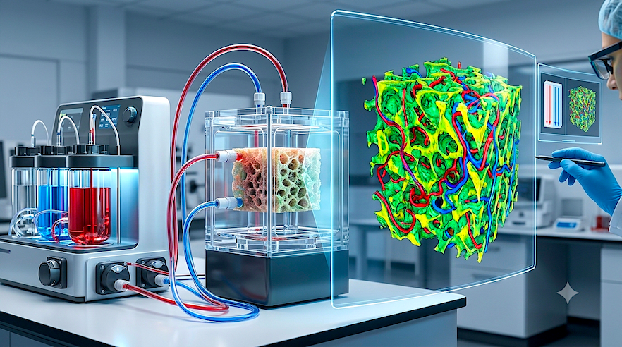
The team’s approach combines custom-designed, 3D-printed components with an Ambr250 bioreactor to maintain tightly regulated culture conditions. The researchers validated the custom parts as autoclavable and leak-proof before using them to support an 8-cm2 macroporous bioprinted tissue. Culture medium was continuously pumped through the tissue at 1.5 mL/min while the bioreactor regulated oxygen levels, temperature, and pH.
To better understand what was happening inside the tissue without damaging it, the researchers used high-resolution magnetic resonance imaging. The scans reconstructed the tissue’s three-dimensional structure and tracked how fluid moved through it. Rather than flowing uniformly, the liquid followed uneven pathways that differed from computer-aided design predictions, revealing a more complex internal environment than expected.
Over a 21-day culture period with this platform, temperature, dissolved oxygen, and pH remained stable across a broad range of oxygen settings. The team did detect a consistent difference between oxygen levels measured in the regulation vessel and those reaching the tissue chamber, but traced the discrepancy to oxygen entering the culture medium through flexible tubing. Still, the researchers successfully perfused three large mesenchymal stem cell–based tissues using minimal culture medium.
Cowles and his colleagues concluded that the new platform provides a controlled environment for cultivating larger bioprinted tissues while offering detailed, non-destructive monitoring of internal flow patterns. Looking ahead, they plan to improve quantitative flow measurements and develop computational fluid dynamics models capable of mapping local tissue microenvironments, with the goal of advancing reproducible large-scale tissue maturation.