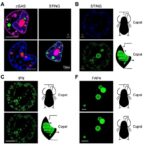
Ras, one of the cell’s most influential molecular switches, has become the focus of a new live-cell imaging technology designed to reveal where and when the protein is active. In a study published in Nature Chemical Biology, researchers introduced HaloRasAR, a chemigenetic biosensor capable of detecting the activity of endogenous Ras in living cells. The system combines a circularly permuted HaloTag protein with the fluorophore JF635, allowing Ras signaling to be visualized with high spatial and temporal resolution. By tracking Ras activity in real time, the researchers aim to address a longstanding challenge in cancer biology: measuring the behavior of native Ras molecules across the diverse cellular compartments where they operate, rather than relying primarily on engineered or overexpressed systems.
Ras proteins are small GTPases that function as molecular switches in pathways controlling cell growth, division, differentiation and survival. They alternate between an inactive state, bound to GDP, and an active state, bound to GTP. When activated, Ras interacts with downstream effector proteins and initiates signaling cascades, including pathways involving RAF kinases, phosphoinositide 3-kinase and other regulators of cell behavior. Mutations that lock Ras in a persistently active configuration are common in human cancers, making Ras a major therapeutic target. Yet Ras activity is not confined to a single location. Although the plasma membrane is a central signaling platform, evidence has shown that Ras can also signal from intracellular membranes, including the Golgi apparatus. This compartmental organization means that a measurement averaged across the whole cell can obscure biologically important differences.
Existing Ras activity reporters have helped establish that signaling is dynamic, but many have limitations when the goal is quantitative analysis of endogenous Ras. Some depend on overexpressed Ras or artificial recruitment systems that can alter the normal balance of signaling components. Others rely on fluorescent protein architectures whose optical properties or expression levels complicate comparisons between cells and experiments. HaloRasAR takes a different approach by using a chemigenetic design. In such systems, a genetically encoded protein component is paired with a synthetic fluorescent ligand. The protein provides targeting and molecular recognition, while the ligand supplies the optical signal. This division can offer improved control over labeling, brightness and experimental timing compared with biosensors that are fluorescent from the moment they are produced.
At the center of HaloRasAR is a circularly permuted HaloTag. Circular permutation reorganizes the order of a protein’s amino acid sequence while preserving its overall fold, creating a new arrangement of the molecular input and output regions. In a biosensor, this structural engineering can make the protein’s fluorescence-related behavior responsive to a biological interaction. HaloTag binds covalently to specially designed synthetic ligands, and in this study the reporter is labeled with JF635, a far-red fluorophore. The resulting fluorescence provides a visible readout of Ras activity in living cells. Because the reporter is intended to operate with endogenous Ras rather than requiring large quantities of an introduced Ras construct, it is positioned as a tool for observing signaling under conditions closer to the native cellular state.
The value of the system lies not simply in producing a fluorescent image, but in connecting fluorescence changes to the geography and timing of Ras signaling. Ras activation can begin rapidly after a receptor on the cell surface detects a growth factor, yet the resulting signal may be transmitted through multiple membrane compartments and regulatory proteins. A reporter that records these changes in live cells can show whether activation is sustained or transient, whether it spreads across the cell or remains localized, and whether different organelles display distinct signaling patterns. The researchers used HaloRasAR to examine Ras activity downstream of growth factor stimulation and protein kinase C activation, two routes that engage cellular signaling networks through different upstream mechanisms. These experiments demonstrated the reporter’s ability to follow Ras dynamics in response to distinct stimuli.
Protein kinase C activation is particularly relevant to the question of signaling location because it can influence pathways that intersect with Ras regulation without simply reproducing the canonical sequence of receptor tyrosine kinase events. By comparing growth factor-driven and protein kinase C-driven responses, the study examined how different inputs shape the spatial behavior of Ras. The biosensor revealed that the activity patterns generated by these stimuli could be followed in living cells, offering a way to distinguish signaling events that might appear similar in a conventional endpoint assay. Such measurements are important because two treatments can produce comparable total Ras activity while engaging different membrane pools, durations or downstream consequences. The ability to resolve these differences may help clarify why cells respond differently to signals that converge on the same molecular switch.
The study also applied HaloRasAR to live-cell pharmacology, using the biosensor to investigate the effects of a Ras(G12C) inhibitor and a Ras guanine nucleotide exchange factor inhibitor. Ras(G12C) inhibitors are designed to target a specific cancer-associated mutation in which glycine at position 12 is replaced by cysteine. These drugs exploit the altered chemical environment created by the cysteine residue to bind the mutant protein and suppress its signaling. GEF inhibitors act further upstream or at the activation interface, interfering with guanine nucleotide exchange factors that promote the transition of Ras from its GDP-bound state to its active GTP-bound state. By imaging inhibitor responses rather than measuring only a final biochemical output, the researchers observed subcellular-specific inhibition profiles, indicating that drug effects can differ depending on where Ras signaling is occurring.
That observation has significant implications for the development and evaluation of targeted therapies. A compound may strongly reduce Ras activity in one cellular compartment while leaving another pool comparatively less affected. Differences may arise from drug access, local protein partners, variations in nucleotide cycling or distinct regulatory mechanisms operating on different membranes. Standard assays that extract and average cellular material can miss this compartmental behavior. HaloRasAR provides a means of watching the response unfold in individual living cells, potentially exposing rapid inhibition, delayed recovery or residual signaling that would otherwise be hidden. This could help researchers understand why some tumors respond incompletely to Ras-directed treatment and how combinations involving Ras, GEFs or downstream pathway components might be optimized.
The new biosensor does not eliminate the complexity of Ras biology, but it offers a platform for investigating that complexity with greater resolution. Its chemigenetic architecture combines the programmability of synthetic fluorophores with the targeting capabilities of genetically encoded sensors, while the use of JF635 supports imaging in the far-red region of the spectrum. That optical range can be useful in experiments involving other fluorescent markers, enabling researchers to monitor Ras activity alongside organelle identities, cell morphology or additional signaling events. Because the reporter is designed for endogenous Ras, it may also reduce some of the interpretive problems associated with overexpression. The authors describe HaloRasAR as a major advance for spatiotemporal interrogation of Ras signaling and live-cell pharmacology, opening the door to more precise studies of how oncogenic and normal Ras activity is organized within cells.
The broader significance of the work extends beyond Ras itself. Many signaling proteins are controlled not only by their biochemical state but also by their location, duration and molecular neighborhood. Tools that can measure these dimensions simultaneously are increasingly important for understanding how diseases emerge and how drugs act inside living cells. HaloRasAR gives researchers a way to ask whether a treatment truly suppresses the relevant Ras pool, whether signaling shifts to another compartment and how cells adapt over time. As Ras-targeted therapies continue to expand, such information could become essential for interpreting resistance, identifying vulnerabilities and designing more effective drug combinations. By turning endogenous Ras activity into a visible, dynamic signal, the technology brings the field closer to watching cancer-associated signaling as it happens rather than reconstructing it after the fact.
Subject of Research: A chemigenetic live-cell biosensor for tracking endogenous Ras activity and evaluating Ras-targeted pharmacology across subcellular compartments.
Article Title: A chemigenetic endogenous Ras activity biosensor for live-cell pharmacology
Article References: Weeks, R., Zhou, D.R., Frei, M.S. et al. A chemigenetic endogenous Ras activity biosensor for live-cell pharmacology. Nature Chemical Biology (2026). https://doi.org/10.1038/s41589-026-02276-0
Image Credits: AI Generated
DOI: https://doi.org/10.1038/s41589-026-02276-0
Keywords: Ras, Ras activity, HaloRasAR, chemigenetic biosensor, HaloTag, JF635, live-cell imaging, cancer signaling, Ras inhibitors, protein kinase C, growth factor signaling, live-cell pharmacology
Tags: advances in molecular imaging of cell signalingcellular compartment-specific Ras activity measurementchemigenetic biosensors for endogenous Rasdrug development targeting Ras pathwaysfluorescent biosensors for cell signalingfluorescent tagging of Ras proteinsGTPase molecular switch monitoringHaloRasAR fluorescence imaginglive-cell imaging of Ras in cancer researchRas activity visualization in live cellsreal-time Ras signaling trackingspatial and temporal resolution of Ras activity