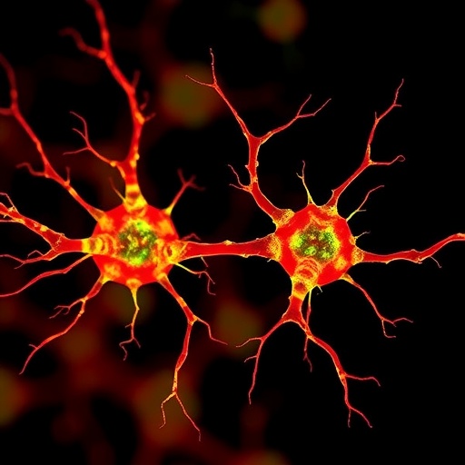
Scientists at the University of Cologne, University Hospital Cologne, and the Max Planck Institute for Metabolism Research have developed a computational method that can measure cellular damage with unprecedented precision. Rather than treating an organ as a uniform mass of healthy or diseased tissue, the approach evaluates the condition of individual cells by examining their patterns of gene activity. The researchers say this could transform how scientists study slowly progressing diseases, including disorders of the kidney and liver, where damage may accumulate over years before symptoms become obvious. Their findings, published in Cell Genomics, introduce cell-type-specific “damage scores” that can reveal how disease develops across individual cells and tissues.
The method is designed to work with two particularly important cell populations: podocytes in the kidney and hepatocytes in the liver. Podocytes are specialized cells that wrap around tiny blood vessels in the kidney’s filtration units, helping prevent essential proteins from leaking into urine. Hepatocytes perform many of the liver’s core functions, including metabolism, detoxification, and the production of blood proteins. Both cell types are vulnerable to age-related and degenerative disease, but they do not necessarily deteriorate at the same speed or through the same biological pathways. By identifying gene-expression signatures associated with cellular injury, the researchers created a way to estimate the degree of damage in each cell rather than assigning a single disease label to an entire tissue sample.
The technique relies on transcriptomics, the large-scale measurement of RNA molecules produced by cells. RNA provides a molecular snapshot of which genes are active at a particular moment. In single-cell RNA sequencing, tissue is separated into individual cells before the RNA inside each cell is measured. This allows researchers to distinguish cell populations and compare their molecular states one by one. Spatial transcriptomics adds another layer of information by recording gene activity while preserving the location of cells within a tissue section. The Cologne-led team developed damage scores that can be applied to both types of data, combining molecular identity with information about the severity of cellular injury and, in the case of spatial data, its position in the tissue.
A central achievement of the work is the identification of marker genes that reflect damage specifically within podocytes and hepatocytes. Because different cell types respond to stress in distinct ways, a general damage signature can obscure important details. A gene that signals injury in a kidney filtration cell may have a completely different meaning in a liver cell. The researchers therefore used a computer-assisted strategy to select patterns that are informative for the cell type being studied. The resulting scores allow cells to be arranged along a continuum, from relatively intact to severely damaged, instead of being placed into a simple healthy-versus-diseased category.
This continuous view could provide insight into the order in which disease-related events occur. In a conventional tissue analysis, early molecular changes may be mixed together with signals from advanced injury, making it difficult to determine which processes initiated the decline and which appeared later. With the new approach, researchers can compare cells at different levels of damage and reconstruct likely disease trajectories. They can ask whether inflammation, metabolic disruption, loss of cellular specialization, or stress responses appear first, and whether those changes are followed by structural damage or cell death. Such information may help identify periods during which diseased cells remain vulnerable but potentially recoverable.
The method may also expose differences between patients that are hidden by conventional averages. Two people may receive the same diagnosis while having very different proportions of mildly, moderately, and severely damaged cells. Their tissues may also rely on different biological pathways as disease progresses. By examining the distribution of damage scores across cells, scientists could distinguish mechanisms shared by many patients from changes that are specific to an individual. This distinction is particularly important for degenerative diseases, which often develop gradually and may not respond uniformly to the same treatment. The researchers suggest that the approach could ultimately support more precise predictions about how a patient’s condition will evolve.
The work emerged from collaboration between computational biologists and clinicians. Andreas Beyer, a professor associated with the Cluster of Excellence on Aging Research CECAD, led the computational side of the study, while Martin Kann, a nephrologist, helped establish the clinical focus on kidney disease. Liver and metabolism specialists later joined the effort, expanding the method beyond a single organ. This combination of expertise enabled the team to connect patterns found in sequencing data with the biological and medical features of tissue injury. It also reflects a growing shift in biomedical research toward integrating laboratory algorithms with samples obtained in clinical settings, including biopsies.
Although the initial applications focus on podocytes and hepatocytes, the researchers say the underlying strategy is not limited to these cells or organs. In principle, the same framework could be adapted by identifying appropriate marker genes for other specialized cell types. That could make it useful for studying diseases in the heart, brain, lung, or intestine, provided researchers can establish reliable molecular signatures of cellular damage. Its compatibility with both single-cell and spatial transcriptomics is another advantage. Single-cell data can provide detailed molecular measurements across thousands of cells, while spatial data can show whether damaged cells cluster near blood vessels, ducts, fibrotic regions, or other structural features.
The scientists are now refining the method to improve its ability to predict disease progression. A more accurate forecast could help researchers identify early intervention points and evaluate whether a treatment is reversing damage, slowing its spread, or merely controlling symptoms. In the longer term, cell-specific damage scores could support the design of clinical studies by helping investigators select patients with comparable disease states or measure treatment effects at a molecular level. The approach does not replace clinical diagnosis, but it may provide a much finer resolution of what is happening inside diseased tissue. By turning gene-expression patterns into measurable trajectories, the study offers a new way to follow disease from its earliest cellular disruptions to its later consequences.
Subject of Research: Human tissue samples
Article Title: Cell-type-specific damage scores reveal kidney and liver disease trajectories in single-cell and spatial transcriptomics
Web References: https://doi.org/10.1016/j.xgen.2026.101337
References: Cell Genomics, DOI: 10.1016/j.xgen.2026.101337
Keywords: single-cell RNA sequencing, spatial transcriptomics, cell damage, kidney disease, liver disease, podocytes, hepatocytes, gene expression, disease progression, computational biology, aging research
Tags: advanced techniques for studying aging-related tissue damagecell-level insights into disease development and progressioncell-type-specific damage scoring in chronic diseasescellular damage assessment in degenerative diseasescomputational methods for measuring cell-specific disease progressiongene activity patterns in podocytes and hepatocyteshigh-precisionimpact of degenerative diseases on individual cell functioninnovative research in kidney and liver disease diagnosticsmolecular signatures of cellular deteriorationpersonalized analysis of cellular degenerationsingle-cell gene expression analysis in kidney and liver cells