Soybean Flavonoids Push Antibiotic-Factory Bacteria Into a Wall-Less State
A pair of compounds found abundantly in soybeans can coax one of the world’s most important antibiotic-producing bacteria into shedding its protective cell wall, researchers report. When exposed to the plant flavonoids daidzein and genistein while under intense osmotic stress, Streptomyces coelicolor transformed from branching filaments into rounded, wall-deficient cells—some of which became capable of dividing. The discovery identifies plant chemicals as direct triggers of an unusual bacterial survival and growth state, offering a new window into how soil microbes respond to their chemical surroundings and potentially creating a controllable route for biotechnology.
The finding is striking because Streptomyces bacteria are not ordinary single-cell organisms. They grow as branching, fungus-like hyphae and pass through a complex multicellular life cycle that includes differentiation, programmed cell death and the production of spores. Their filamentous networks are also biochemical powerhouses: members of the group produce more than two-thirds of clinically used antibiotics, along with anticancer, immunosuppressive and agricultural compounds. Much of this biology depends on the cell’s peptidoglycan wall, a mesh-like polymer that provides mechanical strength and prevents the membrane from bursting. Yet Streptomyces can sometimes abandon this structure and adopt alternative forms known as wall-deficient cells.
The researchers distinguish two important versions of this unusual state. S-cells are non-dividing, stress-induced cells that lack a rigid wall but remain metabolically active and retain the machinery needed to rebuild filamentous growth. L-forms, by contrast, are wall-deficient cells that can reproduce autonomously. Both are fundamentally different from extracellular vesicles, which are membrane-bound particles released by bacteria and generally cannot replicate. S-cells may look superficially similar to vesicles because they are rounded and wall-less, but they are living cells rather than inert packages. L-forms can divide without the conventional FtsZ-based divisome, the protein machinery that normally organizes bacterial cytokinesis and directs the construction of a new septum.
Previous experiments had shown that wall-deficient cells could appear in S. coelicolor after specific genetic changes, particularly when the bacteria were exposed to high concentrations of sucrose. But osmotic protection alone was not enough. At 0.3 molar sucrose, the medium could prevent wall-less cells from bursting, yet it did not reliably initiate their formation. A stronger treatment, 0.64 molar sucrose, was required, and even then the wild-type bacterium did not produce these forms in conventional glucose-yeast extract-malt extract medium. The new study began with an unexpected observation: the same strain readily formed rounded cells in soya flour-mannitol medium under the stronger osmotic challenge. That suggested that a soybean-derived molecule, rather than nutrients in general, might be acting as a morphogenetic signal.
Soybeans are rich in isoflavones, a class of flavonoids that includes daidzein and genistein. These molecules are already known to influence bacterial physiology, alter membrane properties and participate in plant-microbe communication. In legume roots, for example, they help activate gene expression in symbiotic rhizobia, bacteria that invade plant tissues and establish nitrogen-fixing partnerships. The researchers estimated that the soya flour medium contained roughly 21 micrograms per millilitre of daidzein and 22.2 micrograms per millilitre of genistein. They therefore added purified versions of the two compounds to flavonoid-free laboratory medium and tested whether they could reproduce the soybean effect.
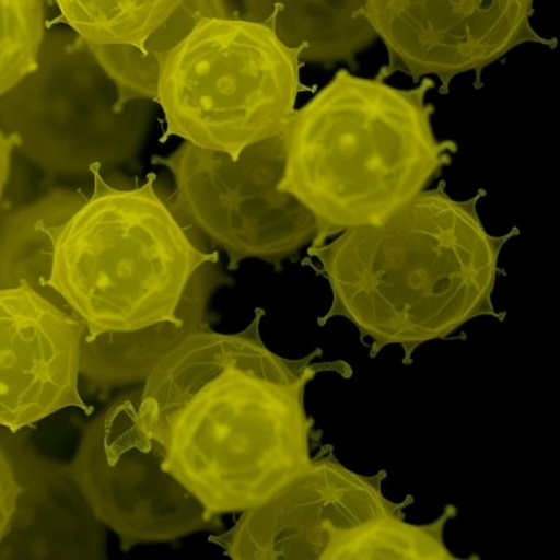
The results were dose-dependent and dramatic. At 6.25 micrograms per millilitre, either flavonoid produced small but detectable numbers of rounded cells. Combining the compounds strengthened the response, generating cells with an average area of 5.9 square micrometres. At 15 micrograms per millilitre, the formation of these structures increased sharply. At 25 micrograms per millilitre—close to the estimated concentration in the soybean medium—the bacteria produced much larger S-cells. When daidzein and genistein were combined at this concentration, the average cell area reached 17.6 square micrometres, and individual structures grew to as much as 35 square micrometres. Under the most effective conditions, nearly all of the stained cellular material adopted the rounded S-cell morphology, leaving virtually no recognizable filamentous hyphae.
Microscopy confirmed that the transformation was not simply a swelling of ordinary hyphae. The team used FM5-95, a fluorescent membrane dye, and wheat germ agglutinin linked to Alexa Fluor 488, a probe that binds the N-acetylglucosamine and N-acetylmuramic acid residues characteristic of peptidoglycan. The induced structures were surrounded by membranes but showed no detectable cell-wall staining, demonstrating that they were genuinely wall-deficient. Some cells retained thin wall fragments or discrete patches of peptidoglycan, possibly marking remnants of the original wall or locations where wall synthesis and degradation were still occurring. A subset also displayed unusually strong internal membrane staining, a feature associated with L-form biology in other bacteria.
Time-lapse imaging revealed that most of the stress-induced cells behaved as non-dividing S-cells, but a small minority crossed into the L-form state. Among 137 cells tracked over 17 hours, nine—about 6 percent—showed pronounced deformation and movement. Three cells, approximately 2.2 percent of the total, moved and divided. The dividing L-forms appeared to establish transient membranous bridges between daughter cells, structures that resemble midbody-like connections described during cytokinesis in animal cells and during L-form division in Listeria monocytogenes. Because wall-deficient division does not depend on the standard septum-building apparatus, such observations may help researchers understand how membranes themselves can generate new cells when the rigid bacterial wall is absent.
The molecular explanation remains unresolved, but the experiments point toward an interaction between flavonoid activity and the machinery that remodels peptidoglycan. The researchers examined mutants affecting four proteins: the N-acetyltransferase SCO0954, the D-alanyl-D-alanine carboxypeptidase SCO4439, the GOLPH3-like protein SCO4440 and the EngA GTPase SCO1758. These proteins are involved directly or indirectly in cell-wall remodeling, and mutations in their genes can promote S-cell and L-form formation under hyperosmotic stress. In soybean medium, the mutant carrying a disruption affecting sco1758 produced significantly smaller and fewer wall-deficient cells, while the sco0954 knockout and a strain overexpressing sco0954 approached complete conversion to S-cells. The pattern suggests that flavonoids do not act through a single isolated switch but may lower the physiological barrier to wall removal while the bacterium’s peptidoglycan and membrane systems determine how far the transformation proceeds.
One possibility is that daidzein and genistein alter membrane fluidity, permeability or redox balance, indirectly destabilizing the coordination between membrane growth and peptidoglycan synthesis. Flavonoids are often discussed as antimicrobial compounds because, at sufficiently high concentrations, they can damage bacterial membranes and disrupt cellular homeostasis. But the new results suggest that their effects can be more nuanced than simple toxicity. At the concentrations tested—roughly 60 to 100 micromolar—the compounds may function as environmental signals in combination with severe osmotic stress, prompting a reversible change in cell architecture. The study examined only two flavonoids, so it is not yet clear whether the response is specific to soybean isoflavones or represents a broader reaction to membrane-active plant phenolics.
The ecological implications are tantalizing but still speculative. Daidzein and genistein are released into the rhizosphere by legume roots, where concentrations can vary sharply across microscopic distances and may become locally elevated at the plant surface. A wall-less state could potentially help soil-dwelling actinomycetes negotiate crowded, chemically complex plant-associated habitats. Wall-deficient bacteria are flexible and deformable, and L-forms in other species have been linked to stress tolerance, antibiotic persistence and resistance to certain bacteriophages. There is also a natural precedent for wall-less bacteria living inside plants: phytoplasmas lack peptidoglycan entirely and inhabit phloem tissues, moving through narrow sieve pores. None of this demonstrates that Streptomyces S-cells colonize plants in nature, however. Establishing that connection will require experiments in soil, on living roots and in other streptomycete species.
The work could also matter for biotechnology. Streptomyces already supplies a vast catalogue of medically valuable molecules, while bacterial extracellular vesicles are being explored as delivery vehicles for proteins, nucleic acids and therapeutics. S-cells and L-forms are not vesicles, but their membrane-rich, wall-deficient architecture could provide a complementary platform for studying or engineering the movement of biological cargo. The flavonoid trigger offers something that has been missing: a defined chemical means of inducing the state without relying solely on genetic mutations or poorly characterized soybean extracts. Before such applications become realistic, researchers will need to determine whether induced cells can be recovered efficiently, how reliably they return to normal filamentous growth, what they contain, and whether the process can be controlled without compromising viability. For now, the central message is that a molecule produced by a plant can push a major bacterial antibiotic producer into a radically different form—revealing just how responsive microbial cell architecture can be to the chemistry of its environment.
Subject of Research: Flavonoid-induced wall-deficient cell formation in Streptomyces coelicolor under hyperosmotic stress
Subject of Research: Biology
Article Title: The Flavonoids Daidzein and Genistein Induce Wall-Deficient Cell Formation in Streptomyces coelicolor Under Hyperosmotic Stress
Article References: Valdés‐Chiara, P., Alonso‐Fernández, S., Manteca, A., & Fernández‐García, G. (2026). The Flavonoids Daidzein and Genistein Induce Wall‐Deficient Cell Formation in Streptomyces coelicolor Under Hyperosmotic Stress. Microbial Biotechnology, 19(6), Article e70366. https://doi.org/10.1111/1751-7915.70366
Image Credits: AI Generated
DOI: 10.1111/1751-7915.70366
Keywords: Streptomyces coelicolor, daidzein, genistein, flavonoids, S-cells, L-forms, hyperosmotic stress, peptidoglycan remodeling, plant-microbe interactions
Cite this news
APA
MLA
Chicago
SCIENMAG. (August 28, 2026). Daidzein and Genistein Trigger Wall-Less Streptomyces coelicolor Cells Under Hyperosmotic Stress. https://scienmag.com/daidzein-and-genistein-trigger-wall-less-streptomyces-coelicolor-cells-under-hyperosmotic-stress/
Copy citation
Download RIS
Tags: antibiotic productionantibiotic-producing bacteria cell wall sheddingbacterial cell wall sheddingbacterial morphological plasticitybacterial response to hyperosmotic stressbiotechnology applicationsbiotechnology applications of wall-deficient bacteriadaidzeinDaidzein and genistein effects on Streptomyces coelicolorfilamentous bacteria differentiationfilamentous bacteria transformation under osmotic stressflavonoidsgenisteinhyperosmotic stressimpact of plant-derived compoundsmicrobial adaptation to environmental stressesmicrobial response to plant chemicalsplant chemicals triggering bacterial survival strategiesregulation of antibiotic biosynthesis in Streptomycessoil microbiome chemical signalingsoybean flavonoids inducing wall-less bacterial cellssoybeansStreptomyces coelicolorwall-less bacterial cells