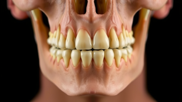
When forensic investigators recover human remains, one of the first and most fundamental questions they must answer is simple to ask but often difficult to answer: was this person male or female? A new study published in the International Journal of Legal Medicine suggests that the answer may lie, quite literally, in the jaw. Researchers from Süleyman Demirel University, Karamanoğlu Mehmetbey University, Lokman Hekim University and Kocaeli City Hospital in Türkiye have demonstrated that the mandible, the largest and strongest bone of the human face, can be used to estimate sex with accuracy rates exceeding ninety percent, using nothing more than three-dimensional reconstructions of routine computed tomography scans.
The study, led by Yadigar Kastamoni and Kenan Öztürk of the Department of Anatomy at Süleyman Demirel University, together with Ahmet Dursun, Onur Can Şanlı and radiologist Veysel Atilla Ayyıldız, set out to reveal sex-related characteristics through morphometric measurements taken on the mandible, and to predict sex in individuals from different age groups using those variables. The work addresses one of the enduring challenges of forensic anthropology: identifying reliable, population-appropriate skeletal markers of sexual dimorphism that remain robust across the adult lifespan.
The research team analysed head and neck computed tomography images from 152 individuals, 77 males and 75 females, aged between 20 and 98 years. From these scans, the researchers generated three-dimensional reconstructions of each mandible using the RadiAnt DICOM viewer, a software platform that converts the two-dimensional slice data produced by CT scanners into volumetric models that can be measured in three dimensions. This approach reflects a broader shift in forensic science away from measurements taken with calipers on dry skulls and toward virtual anthropology, in which clinical imaging archives become rich sources of skeletal data.
The specific landmarks chosen for measurement were carefully selected regions of the mandible with well-established anatomical identities. The team measured dimensions relating to the foramen mentale, the mental foramen through which the mental nerve and vessels emerge on the front of the jaw; the condylus mandibulae, the rounded condyles that articulate with the skull at the temporomandibular joints; the incisura mandibulae, the notch between the condyle and the coronoid process on the upper margin of the ramus; and the angulus mandibulae, the angle of the jaw at the back and lower corner of the bone. In addition, the researchers measured the bigonial distance, the straight-line breadth of the jaw between the two gonion points at the angles on either side. All measurements, except those involving the mandibular angle, were carried out in RadiAnt DICOM Viewer 2020.1.1, and the resulting data were analysed statistically using SPSS 20.0 for Windows.
The results were strikingly consistent with the known biology of the human mandible. When the parameters were compared between the sexes, the mean values of all measurements, with the single exception of the left mandibular angle, were larger in males than in females. This pattern mirrors the influence of testosterone-driven bone growth during puberty, which produces a generally larger, squarer and more robust jaw in men, while hormonal and muscular factors in women tend to produce a jaw with a more obtuse gonial angle and finer proportions. The only measurement that failed to show a statistically significant sex difference was the gonial angle itself on the left side, a finding that reinforces a long-standing caution in the literature: angular measures of the jaw are notoriously variable and are influenced by age-related remodelling, tooth loss and chewing forces.
The degree of sexual dimorphism varied considerably across the measured parameters. The highest dimorphism rate was observed in the incisura mandibulae, the mandibular notch, which differed between the sexes by 14.39 percent on the right side and 14.29 percent on the left. At the opposite extreme, the angulus mandibulae showed almost no dimorphism, with differences of just 0.12 percent on the right and 0.57 percent on the left. This gradient is informative for forensic practice: it suggests that linear distances spanning the ramus, particularly between the mandibular condyle and the gonion, carry far more sex-diagnostic information than the angle of the jaw, which should be treated with caution when sexing fragmentary remains.
Indeed, the statistically significant differences between the sexes were found specifically in the distances from the condylus mandibulae to the gonion on both sides of the jaw, alongside the bigonial distance. These findings give forensic anthropologists a small set of hardy landmarks, robust bony points that survive decomposition and trauma relatively well, from which reliable linear measurements can be taken. Because the condyle and gonion are prominent structures on the ramus, they remain measurable even when the delicate alveolar portion of the jaw bearing the teeth has been damaged or lost, a common scenario in archaeological and forensic contexts.
What elevates the study beyond a simple descriptive anatomy exercise is its predictive modelling. The researchers built a logistic regression model from their measured variables, which correctly classified 91.4 percent of individuals in the development sample. A parallel discriminant function analysis achieved 90.1 percent classification accuracy on the original sample, and, crucially, retained 85.5 percent accuracy under leave-one-out cross-validation, a stringent test in which each individual is classified by a model trained on everyone else. The modest drop between original and cross-validated performance indicates that the model generalises reasonably well rather than simply memorising the training data, an important consideration for any method intended for real casework.
The authors conclude that sex estimation can be successfully performed using external measurements of the mandible, and they highlight the bigonial distance and the gonion-to-condyle measurements as particularly valuable parameters for sex differentiation. They suggest the method may prove useful in anatomy, forensic medicine and anthropology, especially in studies requiring sex estimation. This positions the mandible alongside the pelvis and skull as a first-rank bone for sex estimation, with particular value in contexts where those more classically dimorphic elements are missing. The mandible is also among the densest and most durable bones in the human skeleton, frequently surviving fire, fragmentation and long burial, which makes any validated method for reading sex from it disproportionately useful.
The study also carries broader implications for how forensic science will develop in the coming years. Computed tomography is now routinely performed in hospitals worldwide, and archives of clinical scans represent vast, untapped populations of reference data. Virtual osteometry of this kind does not require a physical skeleton at all: it can be applied ante-mortem to living patients in identification contexts, and post-mortem using clinical or forensic CT, avoiding destructive sampling of evidence. Earlier work cited by the team, including validation studies of three-dimensional CT measurements and postmortem CT morphometry of the mandible in Japanese populations, had already established the technical reliability of imaging-based measurement; the new study extends that framework with a large, wide-ranging adult sample spanning nearly eight decades of age, from 20 to 98 years.
As with all skeletal sex-estimation methods, population specificity remains the central caveat. The dimensions of the mandible vary across ancestral and geographic groups, and the classification functions derived here were developed on a Turkish sample; forensic practitioners applying them elsewhere would need to validate or recalibrate the model against local reference data before relying on it in identification casework. The retrospective design, approved by the Human Research Ethics Committee of Süleyman Demirel University with informed consent waived for anonymised data, also means the findings describe a clinical imaging population rather than a random demographic one. Nevertheless, with cross-validated accuracy approaching ninety percent from a handful of simple linear measurements, the message of the study is clear and, for a field where even a single additional clue can resolve an identification, quietly revolutionary: the jawbone remembers who you were, and it is willing to tell.
Subject of Research: Sex estimation from morphometric measurements of the human mandible in three-dimensional computed tomography images
Article Title: Sex estimation from the mandible in 3D computed tomography images
Article References: Kastamoni, Y., Dursun, A., Şanlı, O. C., Ayyıldız, V. A., & Öztürk, K. (2026). Sex estimation from the mandible in 3D computed tomography images. International Journal of Legal Medicine. https://doi.org/10.1007/s00414-026-04001-x
Image Credits: AI Generated
DOI: 10.1007/s00414-026-04001-x
Keywords: sexual dimorphism, mandible, computed tomography, forensic anthropology, 3D imaging, morphometry, sex estimation, bigonial distance, logistic regression, discriminant analysis, human identification, osteometric analysis
Cite Scienmag News
APA MLA Chicago
Copy citation Download RIS
Tags: 3D CT scan reconstruction3D imagingaccuracy of bone-based sex predictionbigonial distancecomputed tomographydiscriminant analysisforensic anthropologyforensic imaging techniquesforensic radiologyforensic science researchhuman identificationhuman remains identificationjawbone sex estimationlogistic regressionmandiblemandible morphometric analysismorphometryosteometric analysispopulation-specific skeletal markerssex estimationsexual dimorphismsexual dimorphism in human bonesskeletal sex determination