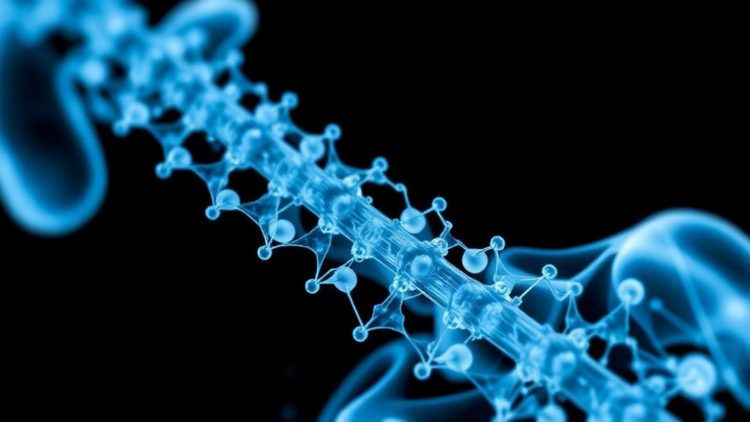
A team of researchers in China has engineered a remarkable new material that could one day change how medicine approaches one of its most devastating conditions: spinal cord injury. Writing in the Journal of Saudi Chemical Society, Tao Xu, Xiaoyu Cai, Weibin Sheng and colleagues at the First Affiliated Hospital of Xinjiang Medical University describe a hybrid nanomaterial that fuses silver nanoparticles with a metal-organic framework, all synthesized using nothing more exotic than an aqueous leaf extract of Tribulus terrestris, a spiny weed better known in traditional medicine circles as puncture vine. When tested in a rat model of contusive spinal cord injury, the compound improved hindlimb function, preserved motor neurons, reduced scar-forming astrocyte activity, and even sharpened sensory recovery. The work, published as an open-access study, sits at the intersection of green chemistry, nanotechnology, and neurotrauma research, and it offers a tantalizing glimpse of how plant-derived nanomaterials might eventually be harnessed to protect the injured nervous system.
The clinical problem the team set out to address is formidable. Spinal cord injury, whether caused by traffic accidents, falls, violence, or sports trauma, triggers a cascade of secondary damage that often proves more destructive than the initial mechanical blow. Within hours of impact, the injured cord becomes a battleground of reactive oxygen species, inflammatory signaling, lipid peroxidation, mitochondrial dysfunction, and progressive cell death. Cavity formation and glial scarring follow, walling off damaged tissue and blocking any prospect of neural regeneration. Conventional management, including early decompressive surgery, corticosteroids, stabilization, and rehabilitation, aims mainly to limit this secondary damage, yet reviews of surgical trials have not consistently demonstrated clear functional gains over conservative approaches. Pharmacological options for the neuropathic pain that often accompanies injury provide only partial relief and carry side effects that can worsen existing deficits. It is precisely this therapeutic vacuum that has pushed researchers toward novel biomaterials capable of intervening in the biochemical storm that follows trauma.
The researchers’ choice of synthesis route is what gives the study its distinctive character. Rather than relying on harsh chemical reducing agents, the team turned to Tribulus terrestris, an annual plant of the Zygophyllaceae family that thrives in arid environments and has long featured in Chinese and Ayurvedic medicine. Fresh leaves collected in the mountains of Chengdu, Sichuan province, were dried, ground, and extracted in sterile double-distilled water. The resulting filtrate, rich in alkaloids, flavonoids, cardenolides, triterpenoids, and steroids, served a dual role in the synthesis: its phytochemicals reduced silver ions to metallic silver nanoparticles and simultaneously capped them, preventing the particles from clumping. In the same reaction vessel, terephthalic acid was deprotonated by triethylamine and coordinated with silver nitrate to build the metal-organic framework in situ, after which the entire composite was coated with hyaluronic acid, a natural polymer prized for its biocompatibility. The whole process ran at ambient temperature, required no toxic solvents beyond the ethanol and dimethylformamide used to dissolve the linker, and yielded a stable suspension in a matter of hours.
Characterization of the resulting material was exhaustive. Fourier-transform infrared spectroscopy revealed the amide band near 1687 per centimeter, carboxylate vibrations of the framework linker at 1569 per centimeter, and a distinctive silver-oxygen stretching band around 526 per centimeter, confirming that plant-derived functional groups had successfully integrated onto the nanoparticle surface. X-ray diffraction displayed the characteristic reflections of face-centered cubic silver at 38, 44, and 64 degrees two-theta, and application of the Debye-Scherrer equation gave a crystallite size of 24.51 nanometers. Energy-dispersive X-ray analysis confirmed metallic silver through strong absorption peaks at 3.1 and 3.2 kiloelectronvolts. Field-emission scanning electron microscopy and transmission electron microscopy showed lumpy, irregularly spherical and oval particles averaging 37.58 nanometers, with some agglomeration attributed to high surface energy and drying artifacts during grid preparation. Perhaps most importantly for biomedical use, the zeta potential measured minus 31 millivolts, a value generally regarded as a hallmark of electrostatic colloidal stability.
Before any animal work began, the team ran a battery of in vitro biological assays. An MTT test against human umbilical vein endothelial cells, a standard model of normal human vasculature, showed minimal cytotoxicity across a wide dose range from 1 to 1000 micrograms per milliliter, an encouraging sign that the composite spares healthy cells. Antioxidant capacity was assessed with the DPPH free-radical scavenging assay, a workhorse technique in which the purple DPPH radical loses color as it is neutralized by electron or hydrogen donation. The nanocomposite achieved an IC50 of 37 micrograms per milliliter, far more potent than the raw leaf extract at 208 micrograms per milliliter, though still short of the synthetic standard BHT at 14 micrograms per milliliter. The researchers attribute this enhanced scavenging to the surface-bound flavonoids and phenolic compounds that concentrate antioxidant chemistry at the nanoparticle surface and facilitate electron transfer.
Antibacterial performance proved equally striking. In agar disk diffusion tests, the composite produced growth inhibition zones against both Staphylococcus aureus and Escherichia coli O157:H7 that in several cases exceeded those of conventional antibiotics including gentamicin, chloramphenicol, and ampicillin. Minimum inhibitory concentrations were 15 milligrams per milliliter for Staphylococcus aureus and 31 milligrams per milliliter for Escherichia coli, with minimum bactericidal concentrations of 31 and 62 milligrams per milliliter respectively. The mechanism, well documented for silver nanoparticles, involves electrostatic binding to negatively charged bacterial membranes, disruption of membrane integrity, penetration into the cell, and damage to DNA, lipids, and proteins, all amplified by the sustained release of silver ions that bind sulfur-containing enzyme residues and trigger lethal reactive oxygen species overload. For spinal cord injury patients, who face elevated risks of infection and infection-associated inflammation that can worsen tissue damage, this antimicrobial activity adds a potentially valuable secondary benefit.
The centerpiece of the study, however, was the in vivo experiment. Forty male Wistar rats, roughly sixty days old, were divided into four groups: intact, sham-operated with saline, spinal cord injury without treatment, and spinal cord injury treated with the nanocomposite. A contusive injury was induced at the T8 level using a weight-drop method, a model chosen because it closely mimics the biomechanics and pathology of human traumatic spinal cord injury, including hemorrhage, ischemia, inflammation, glial scar formation, and progressive cavity development. Beginning one day after injury and continuing daily for two weeks, the treated group received intraperitoneal injections of the nanocomposite at 100 micrograms per kilogram, while the sham group received saline. All procedures were approved by the institutional ethics committee and conducted under established international guidelines for animal experimentation.
The functional results were compelling. Weekly assessment with the Basso, Beattie, and Bresnahan locomotor scale, the standard 21-point measure of rat hindlimb recovery, showed a significant improvement in the treated animals compared with untreated injured controls. Electromyography revealed a markedly higher motor unit recruitment index in both hindlimbs of treated rats, indicating better neuromuscular signaling. A hot-water tail-flick test demonstrated that treated animals responded to painful thermal stimuli with significantly shorter delays, suggesting that the compound helped preserve or restore nociceptive conduction along ascending sensory pathways, a benefit that extends beyond motor recovery into the realm of sensory protection. Histological examination of spinal cord sections stained with hematoxylin and eosin showed substantially smaller lesion cavities and greater numbers of ventral horn motor neurons in the treated group, while immunohistochemical staining for glial fibrillary acidic protein revealed significantly reduced astrogliosis, the reactive astrocyte proliferation that contributes to scar formation and regeneration failure.
Why might a silver nanoparticle-framework hybrid exert neuroprotection? The authors point to a growing body of preclinical literature. Silver nanoparticles have been shown to shift the balance of macrophages in injured neural tissue away from the pro-inflammatory M1 phenotype toward the repair-promoting M2 state, selectively targeting inflammatory cells while scavenging reactive oxygen and nitrogen species. Previous work in rat contusion models found that locally delivered silver nanoparticles in hydrogel reduced demyelination, vacuolization, and lesion size while improving forelimb locomotor scores. Green-synthesized variants have also mitigated Alzheimer’s-like memory deficits in rats by suppressing inflammatory cytokines and activating the Nrf2 antioxidant pathway. The porous framework architecture of the MOF component may further aid drug delivery and sustained release, while the hyaluronic acid coating enhances biocompatibility. The precise mechanism in this spinal cord model remains to be fully elucidated, and the authors are careful to note that more investigation is needed.
The road from a rat model to the clinic is long, and the researchers themselves caution that human application awaits the completion of clinical trial research. Dosing, biodistribution, long-term toxicity of accumulated silver, and the optimal route of administration all remain open questions, and silver nanoparticles are known to display dose-dependent neurotoxicity in some contexts. Nevertheless, the convergence of low cytotoxicity, robust antioxidant and antibacterial activity, colloidal stability, and measurable functional recovery in a clinically relevant injury model makes this green-synthesized composite one of the more intriguing entries in the emerging field of nanoneurotrauma therapy. If subsequent studies confirm and extend these findings, a spiny weed that gardeners curse and traditional healers prize may yet contribute to a genuinely new chapter in the treatment of spinal cord injury.
Subject of Research: Green-synthesized silver nanoparticle-metal-organic framework composites as neuroprotective agents in spinal cord injury
Article Title: Evaluation of the cytotoxicity, antioxidant, antibacterial, and neuroprotective effects of silver nanoparticles combined with metal-organic framework (MOF) synthesized via Tribulus terrestris leaf extract on the contusive model of spinal cord injury in rats
Article References: Xu, T., Cai, X., Yusufu, A., Mamat, M., & Sheng, W. (2026). Evaluation of the cytotoxicity, antioxidant, antibacterial, and neuroprotective effects of silver nanoparticles combined with metal-organic framework (MOF) synthesized via Tribulus terrestris leaf extract on the contusive model of spinal cord injury in rats. Journal of Saudi Chemical Society, 30(4), Article 44. https://doi.org/10.1007/s44442-026-00093-7
Image Credits: AI Generated
DOI: 10.1007/s44442-026-00093-7
Keywords: silver nanoparticles, metal-organic framework, Tribulus terrestris, green synthesis, spinal cord injury, neuroprotection, antioxidant, antibacterial, nanomedicine, BBB score, astrogliosis, Wistar rats
Cite Scienmag News
APA MLA Chicago
Copy citation Download RIS
Tags: antibacterialantioxidantastrogliosisBBB scorebiomaterials for spinal cord injury treatmenteco-friendly nanotechnology in neurotraumagreen chemistry in neuroregenerationgreen synthesisinnovative treatments for spinal cord injuriesmetal-organic frameworkMetal-Organic Frameworks in MedicineNanomedicinenanomedicine for nerve regenerationnanotechnology for spinal cord injuryNeuroprotectionplant-based nanomaterials for nerve repairplant-derived silver nanoparticlessilver nanoparticle healing mechanismssilver nanoparticlessilver nanoparticles for neuroprotectionSpinal Cord Injurytraditional medicine and nanomaterialsTribulus terrestrisWistar rats