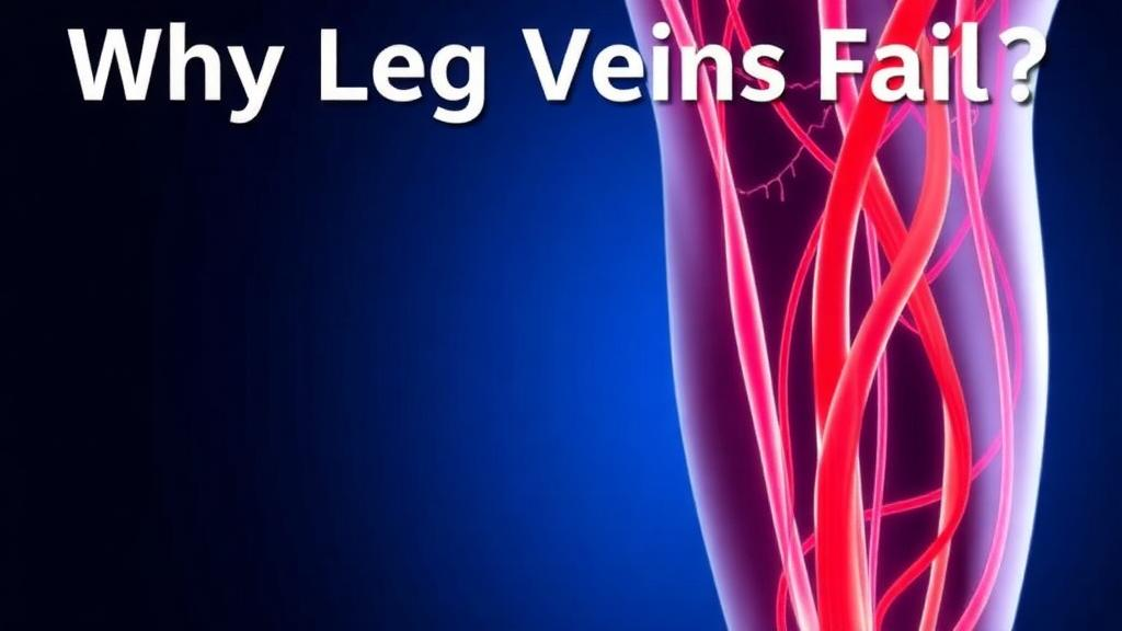
Deep inside the veins of your legs, tiny flaps of tissue are performing one of the most demanding jobs in the human body. Venous valves, thin bicuspid or tricuspid leaflets of connective tissue, open and close roughly two billion times over a lifetime, holding back columns of blood that gravity relentlessly pushes downward. When these valves fail, the result is chronic venous insufficiency, or CVI, a condition that affects an estimated 25 to 40 percent of adults in Western populations, produces symptomatic disease in at least 6 to 7 percent, and costs the United States more than 3 billion dollars annually. Yet despite this enormous clinical burden, the tissue-level mechanics that actually cause a valve to fail have remained almost entirely uncharacterized. A new open-access review published in the Annals of Biomedical Engineering by Nayyan Kaul and Hsiao-Ying Shadow Huang of North Carolina State University now lays out, for the first time in one integrated framework, how experimental measurement, extracellular matrix architecture, mathematical modeling, and computer simulation must fit together to explain venous valve failure.
The review’s central argument is that venous valve biomechanics operates as a strict dependency pipeline, and that a weakness at any stage corrupts everything downstream. Biaxial mechanical testing feeds extracellular matrix characterization; matrix architecture explains mechanical anisotropy; anisotropy determines which constitutive model form is appropriate; and model parameters set the material boundary conditions for fluid-structure interaction, or FSI, simulation. Before 2017, not a single experimentally measured material parameter for venous valve tissue existed. Every computational model of venous valve function published up to that point borrowed linear isotropic stiffness values from arterial tissue or material constants derived from aortic or mitral valve leaflets, structures whose extracellular matrix architecture and loading environments differ from venous valves in ways that no simple scaling factor can correct. The review quantifies just how wrong those borrowed numbers were: fitted constitutive coefficients for venous valve tissue differ from cardiac valve values by factors of 5 to 100 across parameters, meaning the parameter spaces do not even overlap in a way that permits extrapolation.
The experimental foundation of the review rests on biaxial testing, a technique that applies simultaneous tensile loads along two orthogonal axes and captures the coupled mechanical response that uniaxial testing cannot resolve. Venous valve leaflets experience exactly this kind of biaxial loading in vivo: they billow open during forward flow and coapt under retrograde pressure, with the ratio of circumferential to radial stress varying continuously through the cardiac-phasic venous pressure cycle. The only uniaxial study of human tissue, performed in 1985 on femoral vein valve leaflets, established a breaking stress of roughly 11.5 megapascals, about twice that of the adjacent sinus wall, but could say nothing about the coupled deformation behavior that governs leaflet kinematics. Kaul and Huang’s group applied force-controlled biaxial protocols at multiple circumferential-to-radial loading ratios to bovine jugular and saphenous valve leaflets, producing the only systematically characterized nonlinear anisotropic material parameters for venous tissue published to date.
The results revealed a tissue that is strongly anisotropic and highly nonlinear. Jugular valve leaflets reach a peak circumferential tangent modulus of 27 plus or minus 9 megapascals under equibiaxial stretching, versus 9 plus or minus 6 megapascals radially, an anisotropy ratio of approximately 3 to 1 driven by preferential circumferential alignment of collagen fibers. Saphenous leaflets, the tissue directly implicated in lower-limb CVI, are stiffer still, with circumferential peak tangent modulus rising from 62.7 plus or minus 34.9 megapascals proximally to 92.1 plus or minus 27.9 megapascals distally. Strikingly, the two valve types show opposite proximal-to-distal stiffness gradients that track opposing collagen concentration gradients, a crossover that establishes jugular and saphenous valves as mechanically distinct tissues rather than interchangeable surrogates. For comparison, aortic valve leaflets exhibit circumferential moduli of 10 to 15 megapascals with an anisotropy ratio near 10 to 1, ruling out direct cross-tissue material substitution. Intra-valve variability of 30 to 40 percent in tangent modulus further means that single-leaflet sampling systematically underestimates the population-level mechanical range.
Why does venous valve tissue behave this way? The answer lies in its extracellular matrix architecture, which the review treats not as incidental background but as the mechanistic explanation of mechanical behavior. Imaging reveals a clear bi-layer organization: on the parietal, wall-facing side, collagen fibers align preferentially along the circumferential axis with angular deviations of less than 10 degrees in proximal jugular leaflets, resisting the tensile stress that peaks at full valve closure. On the luminal, blood-facing side, a crosslinked radial mesh of elastin dominates, providing the compliance that lets the leaflet billow toward coaptation without tearing. Collagen crimp, the sinusoidal undulation of unstressed fiber bundles, was quantified by second-harmonic generation microscopy at a wavelength of 38.46 plus or minus 8.06 micrometers and an amplitude of 4.51 plus or minus 1.65 micrometers, corresponding to a fiber recruitment threshold of approximately 12 percent true strain. Because physiological circumferential strains during valve closure are estimated at 10 to 20 percent, a substantial fraction of the leaflet’s working range sits precisely within this crimp-recruitment transition, the region where matrix degradation most severely compromises function.
This architecture also explains how disease destroys the valve. Elevated venous pressure triggers an inflammatory cascade that upregulates matrix metalloproteinases, particularly MMP-2 and MMP-9, which selectively degrade fibrillar collagen and elastin. CVI specimens show collagen disorganization and loss of the preferential circumferential alignment that generates the healthy 3-to-1 anisotropy ratio. The consequence is not merely reduced stiffness: the leaflet converts from an anisotropic load-bearing structure into a mechanically isotropic, globally compliant sheet, impairing the directed coaptation force needed for competent closure independent of any change in total collagen content. The authors are careful to note that this interpretation should be read as a plausible proximate mechanical contributor rather than a definitively established causal chain, since direct evidence in human venous valve tissue remains limited, and reported elastin changes in CVI specimens remain contested, likely reflecting differences in disease stage and assay methodology.
On the modeling side, the review identifies a critical failure mode in current constitutive models. The May-Newman and Yin exponential form, fitted to jugular venous data with R-squared values of 0.90 to 0.97 under equibiaxial and near-equibiaxial loading, breaks down systematically under asymmetric conditions. At a 2-to-1 circumferential-to-radial force ratio, the model overpredicts circumferential stiffness and underpredicts radial stiffness, an off-axis prediction error that does not diminish with additional parameter fitting. The mechanism traces to collagen fiber reorientation under off-axis loading, which exponential formulations cannot capture because they lack a structural coupling term to redistribute load across fiber families. The error is most consequential precisely where it matters most: the 2-to-1 ratio at which the model fails coincides with the loading state that dominates physiological valve closure at peak hemodynamic load. The Holzapfel-Gasser-Ogden framework, with its angular fiber dispersion parameter, and the structural constitutive approach of Sacks, which integrates measured fiber orientation distributions directly into the strain energy function, offer principled resolutions, but neither has yet been applied to venous valve tissue.
Three unresolved gaps emerge from the synthesis, each experimentally tractable with existing techniques. First, the off-axis prediction error requires expanded biaxial testing at asymmetric ratios and, ideally, co-registered confocal imaging and mechanical testing on the same specimens, a pairing not yet achieved for venous valves. Second, no failure criterion or damage evolution law exists for venous valve tissue; all biaxial studies to date have operated below the failure threshold, conservatively estimated at 60 to 70 percent true strain, leaving bioprosthetic durability computationally unassessable. Third, no validated material parameters have been published for bioprosthetic candidate materials such as decellularized pericardium, electrospun polyurethane, or silk fibroin scaffolds, even though tissue-engineered venous valves have reached preclinical testing in ovine and porcine models. An isotropic scaffold, the review argues, is mechanically unsuitable as a native surrogate regardless of geometric optimization, because the anisotropy mismatch alone produces divergent closure kinematics and shear distributions.
Perhaps the most sobering caveat is one of species translation: every biaxial dataset currently available derives from bovine tissue, while the clinical target is human. Studies of aortic valves have shown substantial interspecies divergence across humans, pigs, and bovines, and there is no principled reason to assume venous valves behave differently. Human venous valve biaxial characterization remains, in the authors’ framing, the single most consequential missing experiment in the pipeline. The review also flags uncharacterized residual stress, untested viscoelastic effects at physiological loading rates, and an identifiability problem in which multiple parameter sets fit equibiaxial data equally well yet diverge sharply under off-axis prediction. The path forward, the authors conclude, requires no new theoretical frameworks, only disciplined application of established biaxial testing and structural imaging methods to larger and more anatomically diverse specimen sets, ultimately including human tissue. Until then, patient-specific simulation of venous valve function, however sophisticated the software, remains built on a foundation that is only now beginning to be measured.
Subject of Research: Biomechanics, extracellular matrix architecture, and constitutive modeling of venous valve tissue in chronic venous insufficiency
Article Title: From Tissue to Simulation: A Review of Venous Valve Biomechanics, Extracellular Matrix Architecture, and Constitutive Modeling in Chronic Venous Insufficiency
Article References: Kaul, N., & Huang, H.-Y. S. (2026). From Tissue to Simulation: A Review of Venous Valve Biomechanics, Extracellular Matrix Architecture, and Constitutive Modeling in Chronic Venous Insufficiency. Annals of Biomedical Engineering. https://doi.org/10.1007/s10439-026-04412-2
Image Credits: AI Generated
DOI: 10.1007/s10439-026-04412-2
Keywords: chronic venous insufficiency, venous valves, biaxial mechanical testing, extracellular matrix, collagen crimp, elastin, constitutive modeling, fluid-structure interaction, soft tissue biomechanics, tissue-engineered venous valves, matrix metalloproteinases, hyperelasticity