
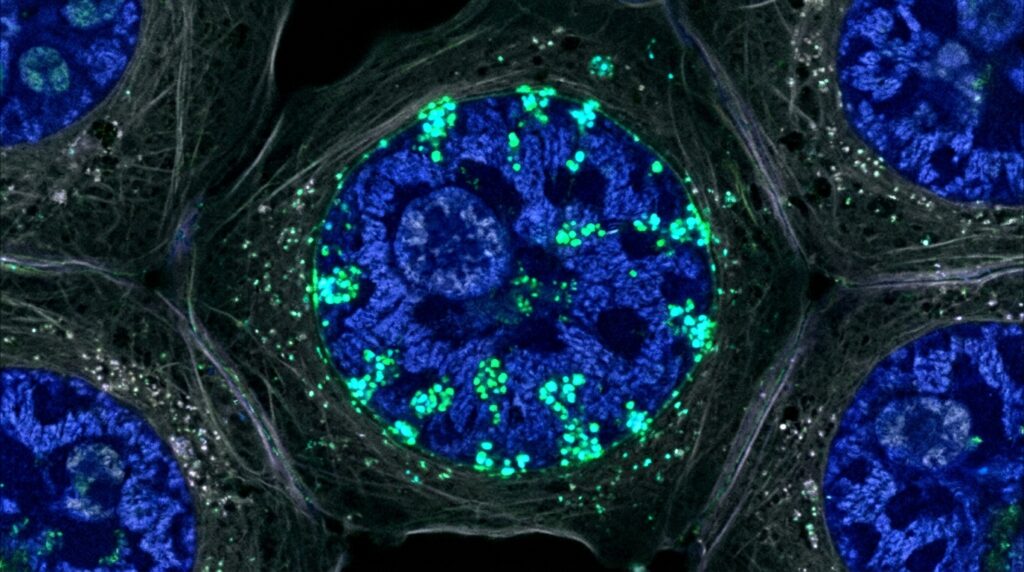
A newly published investigation in Drosophila melanogaster demonstrates that the atypical cadherin Fat functions beyond its established position at the cell membrane by releasing an intracellular fragment that translocates into the nucleus to directly modulate gene transcription. Historically classified as a giant cell adhesion molecule composed of thirty-four cadherin repeats in the extracellular domain, a single transmembrane domain, and an intracellular region, Fat has long been characterized as an apical-junctional regulator of tissue growth, planar cell polarity, and cellular metabolism. In standard models of tissue growth control, membrane-bound Fat acts upstream of the Hippo signaling cascade by governing the kinases Hippo and Warts, which in turn restrict the nuclear translocation and transcriptional activity of the co-activator Yorkie and its DNA-binding partner Scalloped. The present research establishes that the Fat intracellular domain, designated as FtICD, possesses two evolutionarily conserved nuclear localization signals alongside a nuclear export signal. Following an intracellular proteolytic cleavage event, an FtICD fragment dissociates from the plasma membrane and enters the cell nucleus both in cultured cells and in living developing tissues, where it associates directly with chromatin to govern transcriptional programs.
To investigate whether the intracellular region of Fat could enter the nuclear compartment, the researchers expressed a hemagglutinin-tagged Fat intracellular domain construct, FtICD-HA, in cultured Drosophila S2 cells. Confocal immunofluorescence microscopy counterstained with the nuclear envelope marker lamin and DNA stains revealed that the approximately fifty-eight kilodalton FtICD-HA peptide significantly accumulated within nuclei rather than remaining dispersed in the cytoplasm. Because passive diffusion through nuclear pore complexes is restricted for proteins of this molecular weight, and computational prediction tools failed to detect canonical signal sequences, the team systematically generated a series of deletion and truncation constructs transfected into S2 cells. Immunofluorescence analysis categorized these variants into nuclear-enriched, nuclear-excluded, and ubiquitous distribution patterns, delineating two distinct nuclear localization signals and one nuclear export signal. Notably, the first nuclear localization sequence maps within Conserved Region B, while the second coincides with Conserved Region D, both exhibiting strong evolutionary conservation with human Fat4. In contrast, the identified nuclear export sequence overlaps with previously mapped domains known to participate in mitochondrial import and Hippo pathway regulation, indicating that regulatory trafficking sequences within the intracellular domain may direct alternative subcellular fates.
The study verified nuclear translocation in vivo by expressing HA-tagged Fat constructs in third-instar larval eye imaginal discs under the control of the GMR-Gal4 driver, which initiates expression at the morphogenetic furrow. Immediately posterior to the furrow, FtICD-HA remained cytoplasmic, but as cells advanced further away from the furrow, the protein accumulated inside the nuclei of undifferentiated cells, as identified by Bar staining. High-resolution confocal co-staining demonstrated that nuclear FtICD-HA co-localized extensively with propidium iodide-stained genomic DNA while being completely excluded from nucleoli marked by fibrillarin. In striking contrast, overexpressed full-length Fat remained strictly confined to the apical membrane, whereas an engineered construct lacking the majority of the extracellular cadherin repeats while retaining the endogenous transmembrane and intracellular domains, termed FtΔECD, localized throughout the plasma membrane without entering nuclei. Furthermore, when endogenous polytene chromosomes from larval salivary glands were probed with specific anti-Fat antibodies, distinct banded signals were detected along wild-type chromosomes but were entirely absent in ftx13 null mutants. Several of these endogenous Fat polytene bands co-localized directly with RNA polymerase II phosphorylated at Serine 2, providing cytological evidence that endogenous Fat fragments interact with transcriptionally active chromatin loci.
Because nuclear translocation from a transmembrane precursor requires physical severance from the plasma membrane, the researchers developed an in vivo cleavage reporter assay to monitor whether membrane-anchored Fat undergoes proteolytic release. They generated transgenic flies expressing the membrane-tethered FtΔECD construct fused at its C-terminus to the DNA-binding and transactivation domain of Gal4VP16, driven ubiquitously by the tubulin promoter. When these tub-FtΔECD-Gal4VP16 animals were crossed to a reporter line carrying UAS-mCherry, proteolytic cleavage of the fusion protein released the Gal4VP16 moiety to enter the nucleus and drive mCherry transcription. In eye-antennal and wing imaginal discs, mCherry expression appeared in a scattered, salt-and-pepper pattern, demonstrating that membrane cleavage occurs broadly across developing epithelia without strict spatial restriction. In the larval optic lobe, however, reporter activation showed pronounced spatial and temporal regulation, exhibiting strongest mCherry fluorescence in lamina neurons and medulla neuroblasts marked by Deadpan, both of which arise from the neuroepithelium. Given that endogenously tagged Ft-GFP expression is most abundant within the neuroepithelium, this elevated reporter signal indicates that proteolytic processing of Fat is enhanced within proliferative neuroepithelial progenitors and inherited by their differentiated neuronal and neuroblast progeny.
To evaluate the biological activity of nuclear Fat fragments in developing organs, the investigators ubiquitously expressed untethered FtICD in wing discs using the ubi-Gal4 driver and assessed morphological parameters in adult females. Ectopic FtICD expression caused a significant reduction in overall wing area compared to controls (adjusted P < 0.001, n = 16 versus n = 14), alongside an increase in wing roundness (adjusted P < 0.0001) and a marked decrease in crossvein distance both in absolute terms (adjusted P < 0.0001) and relative to wing length (adjusted P < 0.0001). Targeted overexpression using the wing-specific nub-Gal4 driver similarly induced significant alterations in crossvein distance (adjusted P < 0.0001, n = 12) and wing roundness (adjusted P < 0.0001, n = 12), recapitulating classic planar cell polarity defects observed in ft loss-of-function mutants. In the adult compound eye, ubiquitous FtICD expression generated ectopic mini-equators and inverted ommatidia characterized by strict dorsal-ventral polarity inversions, distinct from the rotational phenotypes typical of canonical Hippo pathway disruption. Notably, posterior compartment overexpression of FtICD-3xFLAG using en-Gal4 did not deplete the atypical myosin Dachs from the apical cell cortex, where membrane Fat normally acts, but instead caused an accumulation of Dachs near basal membranes, confirming that FtICD alters organ development through downstream mechanisms independent of apical Dachs depletion.
The researchers next examined whether FtICD directly modulates Hippo pathway target gene enhancers in vivo, focusing on the well-characterized 32-base-pair Hippo Response Element within the death-associated inhibitor of apoptosis 1 (Diap1) locus. Under standard conditions, wild-type HRE drives lacZ transcription in hpo mutant mosaic clones generated by MARCM in response to unphosphorylated, nuclear Yorkie, whereas an enhancer containing mutated Scalloped binding sites, termed HREm6, remains completely inactive. Remarkably, when UAS-FtICD-3xFLAG was co-expressed within hpo42-47 mutant clones in wing discs, the mutated HREm6 reporter was activated, demonstrating that FtICD can stimulate transcription from this Hippo response element independently of intact Scalloped binding motifs. Expression of FtICD in wild-type tissue without hpo mutation failed to activate either HRE or HREm6, indicating that FtICD-driven transcriptional activation still requires Yorkie nuclear localization mediated by reduced Hippo kinase activity. Furthermore, this activation displayed strict tissue selectivity: while FtICD stimulated HREm6 within wing disc clones, it was unable to activate HREm6 in larval eye disc clones, and expression of Expanded, another canonical Hippo target, remained unaffected across the anterior-posterior compartment boundary.
To map chromatin binding across the genome, the team performed chromatin immunoprecipitation coupled with high-throughput sequencing (ChIP-seq) on third-instar larval eye-brain complexes expressing FtICD-3xFLAG driven by ubi-Gal4, employing a dual crosslinking protocol combining ethylene glycol bis(succinimidyl succinate) and formaldehyde. High-confidence FtICD peaks, identified by fold enrichment greater than 2 over controls across biological triplicates, mapped predominantly to gene promoter and enhancer regions. Multiple robust ChIP-seq peaks mapped directly to regulatory elements of the Diap1 locus, including the specific HRE region located upstream of the diap1-RC transcript isoform. Gene Ontology analyses of FtICD-occupied loci revealed substantial enrichment for genes associated with planar polarity, Hippo signaling, chromatin organization, and pattern formation. When cross-referenced with publicly available ChIP-seq datasets from larval eye discs, genome-wide correlation analysis revealed substantial occupancy overlap between FtICD and the canonical Hippo effectors Scalloped (Spearman correlation = 0.56) and Yorkie (Spearman correlation = 0.53). Transcription factor occupancy profiling revealed three major classes of genomic binding: loci occupied preferentially by FtICD alone, loci co-occupied by FtICD together with Yorkie or Scalloped, and regions bound predominantly by Yorkie without FtICD.
To determine the biochemical mechanism allowing a protein lacking recognizable DNA-binding domains to associate with chromatin, the team tested whether FtICD physically interacts with Yorkie and Scalloped in cultured Drosophila S2 cells. Co-immunoprecipitation experiments using anti-FLAG beads revealed that epitope-tagged FtICD-3xFLAG formed a robust physical complex with HA-tagged Yorkie. While full-length Scalloped and its N-terminal half failed to exhibit significant interaction with FtICD, the isolated C-terminal fragment of Scalloped showed strong, reproducible co-immunoprecipitation with FtICD-3xFLAG. Furthermore, an independent yeast two-hybrid screen utilizing an FtICD bait spanning amino acids 4776 to 4998 confirmed a direct physical interaction with a Scalloped prey fragment spanning amino acids 46 to 146. Additional chromatin profiling demonstrated that a distinct subset of FtICD peaks co-bound the transcriptional co-repressor Atrophin and the chromatin factor GAF/Trl, reflecting a separate repressive regulatory module. RNA sequencing of larval eye-brain complexes ubiquitously overexpressing FtICD revealed significant transcriptional upregulation of Hippo pathway components including yorkie (P < 0.0001, 71.05 ± 1.810 transcripts per million), Connector of kinase to AP-1 (P = 0.0093, 127.4 ± 21.23 TPM), Tondu-domain-containing growth inhibitor (P = 0.045, 5.557 ± 1.687 TPM), and Leash (P = 0.0070, 16.54 ± 2.497 TPM), whereas expanded transcription showed no significant alteration (P = 0.2539, -2.488 ± 1.768 TPM).
Beyond the Hippo pathway, transcriptomic profiling revealed bidirectional regulatory activity at diverse non-Hippo genomic loci occupied by FtICD in larval eye-brain tissues. Expression of ATP-dependent chromatin assembly factor large subunit (Acf) was significantly downregulated upon FtICD overexpression (P = 0.0075, -26.88 ± 4.150 TPM), as was partner of numb (pon) (P = 0.0011, -24.21 ± 1.941 TPM). Conversely, several other bound loci showed marked transcriptional upregulation, including CG32581 (P < 0.0001, 87.68 ± 2.329 TPM), Ubiquinol-cytochrome c reductase core protein 2 (P = 0.0206, 183.6 ± 40.90 TPM), rasputin (P = 0.018, 188.9 ± 40.00 TPM), and Kinesin heavy chain (P = 0.0164, 51.93 ± 10.63 TPM). To confirm that chromatin occupancy was not an artifact of ectopic overexpression, the investigators conducted ChIP-seq on 16-to-20-hour embryos using two fertile, homozygous-viable endogenously tagged alleles: Ft-GFPC, harboring a C-terminal GFP fusion, and Ft-GFPint, carrying an internal GFP insertion within the cytoplasmic Hippo N domain at amino acid 4810. After subtracting thirteen nonspecific background peaks identified in w1118 wild-type embryos, the analysis identified 1471 reproducible high-confidence peaks for Ft-GFPC and 1358 for Ft-GFPint, displaying an exceptionally high genome-wide correlation between the two distinct tag locations (Spearman rho = 0.93).
Embryonic ChIP-seq revealed 715 peaks that precisely overlapped between endogenous Fat-GFP lines and ubiquitously overexpressed embryonic FtICD-FLAG, with shared targets including Map205, bazooka, hat-trick, and Ataxin7. Parallel RNA-seq profiling of ft null transheterozygous mutant embryos identified 934 differentially expressed genes, yet intersection with the 791 high-confidence endogenous Fat peak regions revealed that only 19 overlapping genes were significantly altered in expression during embryogenesis, including hat-trick (downregulated by -1.90-fold) and Map205 (upregulated by 2.47-fold). Key Hippo pathway components, such as kibra, diap1, and expanded, showed no significant expression differences in ft mutant embryos, contrasting sharply with their robust transcriptional sensitivity during larval development. De novo motif enrichment analysis within 150 base pairs of endogenous embryonic Fat peak summits revealed significant enrichment for DNA binding sites corresponding to DREF, ZIPIC, M1BP, GATAe, and Homothorax, rather than canonical Hippo elements alone. The authors conclude that membrane-bound Fat releases an intracellular domain fragment that enters the nucleus to associate with Yorkie, Scalloped, and alternative transcription factors in a context-dependent manner, functioning as a nuclear transcriptional regulator to fine-tune developmental signaling.
Subject of Research: Nuclear translocation and transcriptional regulation by the Fat cadherin intracellular domain
Article Title: Fat cadherin cleavage releases a nuclear fragment to regulate gene transcription
Article References: Rusch, J., Thanintorn, N., Kuok, C., Tsatskis, Y., Hou, H., Julick, C., Wilson, M. D., & McNeill, H. (2026). Fat cadherin cleavage releases a nuclear fragment to regulate gene transcription. EMBO Reports. https://doi.org/10.1038/s44319-026-00988-x
Image Credits: AI Generated
DOI: 10.1038/s44319-026-00988-x
Keywords: Fat cadherin, FtICD, Hippo signaling pathway, Yorkie, Scalloped, Planar cell polarity, Proteolytic cleavage, Nuclear localization signals, Chromatin immunoprecipitation, Gene transcription, Drosophila melanogaster, Diap1, Atrophin, Transcriptional regulation, Eye imaginal disc, Wing development, Endogenous ChIP-seq