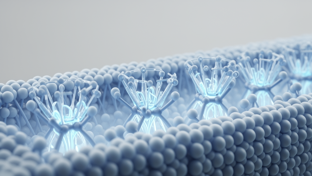
In an investigation into the cellular mechanics of non-invasive therapies for diabetic complications, researchers have demonstrated that negative pressure applied by vacuum erection devices restores erectile function in diabetic rats by activating a specific mechanosensitive signaling pathway. The study, conducted by investigators at Nanfang Hospital of Southern Medical University and Foresea Life Insurance Guangzhou General Hospital, revealed that physical force exerted by the device stimulates the mechanically gated ion channel Piezo1 within the corpus cavernosum. This mechanosensitive activation drives an intracellular influx of calcium and promotes calcineurin-dependent dephosphorylation and nuclear translocation of the transcription factor nuclear factor of activated T cells cytoplasmic 1, commonly designated NFATc1. Across coordinated animal models and primary cell cultures, this mechanotransduction cascade preserved smooth muscle architecture, sustained cellular contractility, and suppressed both apoptosis and fibrotic remodeling within penile erectile tissues.
Diabetes mellitus-induced erectile dysfunction represents a widespread and clinically challenging microvascular complication that exhibits an incidence approximately three times higher in diabetic men than in the general population, affecting up to seventy-five percent of individuals diagnosed with diabetes. First-line pharmacotherapies relying on phosphodiesterase type 5 inhibitors frequently demonstrate markedly lower clinical response rates in patients with diabetic erectile dysfunction compared to those with non-diabetic conditions. This therapeutic shortfall stems primarily from severe structural and functional alterations within the penile corpora cavernosa, characterized by extensive smooth muscle atrophy, persistent apoptosis, heightened oxidative stress, and progressive cavernous fibrosis that extend well beyond impairments in the classic nitric oxide and cyclic guanosine monophosphate signaling pathway. While vacuum erection devices represent an established, guideline-endorsed physical intervention recommended by clinical organizations such as the European Association of Urology, previous scientific inquiries have remained largely confined to evaluating gross hemodynamic outcomes rather than clarifying how controlled external negative pressure is converted into intracellular protective biological signals.
To evaluate these underlying biological mechanisms in vivo, the research team established an experimental model utilizing forty four-week-old male Sprague-Dawley rats. Following overnight fasting, the animals received an intraperitoneal injection of streptozotocin at a dosage of sixty milligrams per kilogram dissolved in citrate-phosphate buffer at pH 4.5 to induce type 1 diabetes mellitus, whereas healthy control animals received equivalent volumes of buffer alone. Persistent hyperglycemia was validated by fasting blood glucose concentrations exceeding 16.7 millimoles per liter on both day three and day seven post-injection. Eight weeks following diabetes induction, diabetic rats underwent functional screening using an apomorphine test administered subcutaneously at eighty micrograms per kilogram to identify individuals exhibiting pronounced erectile dysfunction for subsequent intervention groups. Throughout the entire subsequent experimental timeframe, elevated fasting blood glucose concentrations remained unchanged across all diabetic groups regardless of whether therapeutic intervention was applied.
The animal experiments proceeded across two sequential cohorts to systematically assess therapeutic efficacy and pharmacological modulation. The first cohort evaluated fifteen rats divided into three equal groups consisting of non-diabetic controls, untreated diabetic rats with erectile dysfunction, and diabetic rats receiving vacuum device intervention. The second cohort expanded the evaluation to twenty-five rats distributed across five groups: control animals, untreated diabetic animals, vacuum-treated diabetic animals, diabetic animals treated with the selective Piezo1 channel agonist Yoda1, and diabetic animals receiving vacuum therapy in combination with the mechanosensitive channel inhibitor GsMTx4. Intracavernosal injections of Yoda1 were delivered at a concentration of thirty micromolar in fifty-microliter volumes once daily, five days per week for two weeks. Concurrently, GsMTx4 was administered at ten micromolar using an identical intracavernosal injection schedule immediately following physical vacuum procedures, with mupirocin ointment applied post-intervention to prevent local surface infection.
The physical vacuum protocol utilized a custom cylinder apparatus checked for airtight integrity, applied after lubricating the rat penis and cylinder interior with paraffin oil under isoflurane anesthesia. A negative pressure of negative two hundred millimeters of mercury was established and maintained continuously for five minutes per session, administered five days per week over a total duration of two weeks. Functional erectile recovery was assessed in anesthetized rats by cannulating the left carotid artery for continuous mean arterial pressure monitoring while simultaneously inserting a heparinized saline-filled twenty-four-gauge needle into the mid-shaft corpus cavernosum to record intracavernosal pressure during bipolar electrical stimulation of the cavernous nerves at fifteen hertz, 1.2 milliseconds, and five volts. The ratio of the change in intracavernosal pressure to mean arterial pressure demonstrated severe erectile impairment in untreated diabetic animals, which was markedly reversed by vacuum erection device therapy and significantly recapitulated by Yoda1 monotherapy, whereas GsMTx4 co-treatment partially blunted these functional improvements.
Histological and molecular examination of harvested cavernosal tissues further delineated the structural consequences of the therapeutic interventions. Untreated diabetic animals displayed significant reductions in total corpus cavernosum weight along with extensive apoptotic cell death detected by terminal deoxynucleotidyl transferase dUTP nick end labeling, alongside pronounced collagen deposition and loss of smooth muscle content quantified through Masson trichrome and hematoxylin and eosin staining. Daily application of the vacuum erection device significantly attenuated tissue apoptosis, preserved smooth muscle content, and suppressed collagen accumulation. Western blot and immunohistochemical analyses demonstrated that Piezo1 channel expression, which was drastically downregulated in untreated diabetic penile tissue, was robustly restored following vacuum intervention. Pharmacological activation of Piezo1 via Yoda1 alone partially improved structural tissue architecture and curtailed apoptosis, although its capacity to restore the quantitative smooth muscle-to-collagen ratio was milder than that achieved by whole vacuum mechanical therapy.
To isolate and interrogate the cellular mechanotransduction machinery directly, the investigators isolated primary corpus cavernosum smooth muscle cells from male Sprague-Dawley rats aged six to eight weeks. Primary cells migrated outward from minced cavernosal tissue fragments by day seven of culture, exhibiting characteristic swirl-like growth patterns and double-positive immunofluorescence staining for alpha-smooth muscle actin and calponin. To recreate the mechanical forces generated by the vacuum device in vitro, primary smooth muscle cells seeded on collagen-precoated flexible plates were subjected to twenty percent cyclic equibiaxial mechanical stretch at a frequency of 0.3 hertz for twenty-four hours using a Flexcell tension system. Unstretched static cultures served as baseline controls. RNA sequencing conducted on cells treated with Yoda1 identified NFATc1 as the most significantly enriched transcription factor across multiple pathways, including extracellular matrix-receptor interactions, calcium signaling, and mitogen-activated protein kinase pathways.
Dynamic calcium imaging utilizing the fluorescent probe Fluo-4 acetoxymethyl ester revealed distinct intracellular calcium kinetics governing this mechanotransduction cascade. Direct administration of twenty micromolar Yoda1 triggered an instantaneous spike in intracellular calcium fluorescence, whereas smooth muscle cells subjected to twenty-four hours of cyclic mechanical stretch maintained a sustained, elevated baseline calcium level throughout recording periods. Pre-incubation with the mechanosensitive inhibitor GsMTx4 completely prevented these dynamic calcium responses. Furthermore, three-dimensional collagen gel contraction assays demonstrated that both cyclic mechanical stretching and Yoda1 exposure markedly enhanced the contractile capacity of primary cavernosal smooth muscle cells over twenty-four hours, whereas GsMTx4 counteracted these contractility gains. In harvested tissue specimens, vacuum therapy significantly elevated overall tissue calcium content and increased calcineurin protein concentrations measured by enzyme-linked immunosorbent assay, corroborating downstream calcium-dependent signaling activation in vivo.
The functional dependence of this protective program on downstream transcription factors was resolved through targeted small interfering RNA knockdown experiments in cultured cavernosal cells. Silencing Piezo1 or NFATc1 expression markedly increased basal apoptosis rates as quantified by Annexin V and propidium iodide flow cytometry, accompanied by decreased anti-apoptotic BCL-2 expression, elevated pro-apoptotic BAX, and heightened cleavage of caspase-3. Concurrently, loss of either Piezo1 or NFATc1 elevated fibrotic markers, causing significant increases in Collagen I, transforming growth factor-beta, and phosphorylated Smad2/3 protein levels. Nuclear-cytoplasmic fractionation demonstrated that Yoda1 stimulation and mechanical stretch promoted the dephosphorylation of NFATc1 and facilitated its translocation into the nucleus. When NFATc1 was knocked down, the anti-apoptotic, anti-fibrotic, and contractility-preserving benefits typically elicited by Piezo1 activation were substantially abolished, establishing NFATc1 as the indispensable downstream transcriptional mediator.
The authors highlighted several methodological considerations and boundaries regarding the scope of their findings. First, the experimental investigations focused exclusively on cavernosal smooth muscle cells without examining endothelial Piezo1 expression or activity, leaving open the possibility that endothelial mechanotransduction provides complementary vascular effects during negative pressure therapy. Second, the animal model relied on streptozotocin induction to simulate type 1 diabetes mellitus, whereas the vast majority of clinical diabetic erectile dysfunction occurs in patients presenting with type 2 diabetes accompanied by obesity and insulin resistance. Third, all cellular and molecular analyses were conducted using rodent-derived cells and tissues, necessitating subsequent validation in human primary corpus cavernosum smooth muscle cells to account for potential interspecies differences in mechanosensitive channel kinetics. Finally, the authors observed that chemical stimulation via Yoda1 did not achieve the full magnitude of structural repair provided by mechanical vacuum therapy, underscoring that while Piezo1 activation forms a central therapeutic axis, physical negative pressure likely engages additional parallel mechanobiological mechanisms in the penile corpora cavernosa.
Subject of Research: Mechanotransduction pathway of vacuum erection device therapy in diabetic erectile dysfunction
Article Title: Vacuum erection devices ameliorate erectile dysfunction in diabetic rats via Piezo1/NFATc1-mediated mechanotransduction
Article References: Fan, Y., Gao, G., Pan, M., Liu, Y., Xiang, Y., He, S., Wei, A., & Zhang, H. (2026). Vacuum erection devices ameliorate erectile dysfunction in diabetic rats via Piezo1/NFATc1-mediated mechanotransduction. Frontiers in Pharmacology, 17, Article 2004480. https://doi.org/10.3389/fphar.2026.2004480
Image Credits: AI Generated
DOI: 10.3389/fphar.2026.2004480
Keywords: Vacuum erection device, Erectile dysfunction, Diabetes mellitus, Piezo1, NFATc1, Mechanotransduction, Corpus cavernosum, Smooth muscle cells, Calcineurin, Calcium signaling, Apoptosis, Fibrosis, Yoda1, GsMTx4, Cell contractility, Streptozotocin