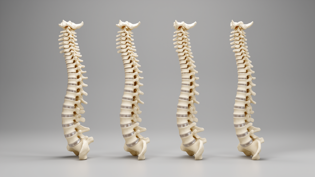
The morphology of the adult cervical spine clusters into four distinct archetypes governed by two uncorrelated dimensions: overall vertebral size and overall spinal canal size. Researchers identified these structural categories through automatic three-dimensional measurements extracted from computed tomography images, providing an empirical framework to describe anatomical diversity across adult populations.
Statistical evaluations showed substantial sex-related variation in spinal structure, with male subjects presenting larger vertebrae than female subjects (p = 6 × 10⁻⁷⁴).
Age also influenced cervical dimensions, with subjects older than 65 years old presenting slightly larger vertebrae (p = 3 × 10⁻²) as well as thinner intervertebral discs (p = 8 × 10⁻³).
In contrast to vertebral size, spinal canal dimensions varied independently of sex and age, meaning that subjects of any demographic profile can present an inherently narrower or wider spinal canal. The authors observed that an inherently narrower spinal canal could carry clinical implications for older individuals as degenerative stenosis progresses.
The study directly addressed an ongoing challenge in biomechanical modeling, where simulations often fail to represent an intended population because spine models are commonly trimmed down to one average or specific subject. Developing a catalogue of archetypical spine morphologies provides a path toward achieving better population representation in engineering and biomechanical simulations.
To establish the catalogue, the researchers assembled a cohort of 450 subjects from publicly available datasets of computed tomography scans originating from the United States, Canada, Switzerland, and Germany. The investigators constructed vertebral templates with identified anatomical landmarks from a 50th-centile subject and elastically registered those templates to the vertebrae of each subject in the dataset.
Using coordinates from the registered landmarks, the team extracted detailed measurements including vertebral body height, width, and depth; spinal canal width and depth; pedicle width, height, and transverse angle; and intervertebral disc height. Hierarchical agglomerative clustering on principal components was then used to analyze the dataset, yielding archetype measurements that institutions can employ as morphing targets to improve the generalizability of simulation results, while providing an automatic measurement framework that can be extended to other organs.
Subject of Research: Cervical spine morphometrics and morphological archetypes derived from computed tomography scans
Article Title: Creation of a Catalogue of Adult Cervical Spine Morphometrics from Automatic Three-Dimensional Measurements of CT-Scan Images and Cluster Analysis
Article References: Diotalevi, L., Clin, J., Hachem, B., LeNaveaux, F., Mac-Thiong, J.-M., & Petit, Y. (2026). Creation of a Catalogue of Adult Cervical Spine Morphometrics from Automatic Three-Dimensional Measurements of CT-Scan Images and Cluster Analysis. Annals of Biomedical Engineering. https://doi.org/10.1007/s10439-026-04446-6
Image Credits: AI Generated
DOI: 10.1007/s10439-026-04446-6
Keywords: cervical spine, spine morphometrics, computed tomography, cluster analysis, biomechanical simulation, vertebral body, spinal canal, degenerative stenosis, intervertebral disc height, pedicle anatomy, elastic registration, anatomical landmarks, principal component analysis, vertebral templates, population representation, hierarchical agglomerative clustering, morphological archetypes