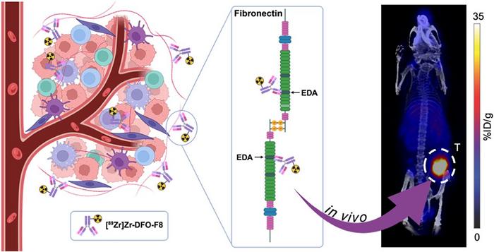
![[89Zr]Zr-DFO-F8 binds specifically with EDA-FN in the tumor stroma to delineate TNBC.](https://bioengineer.org/wp-content/plugins/wp-fastest-cache-premium/pro/images/blank.gif)
A groundbreaking advance in molecular imaging has emerged that promises to transform the diagnosis and management of triple-negative breast cancer (TNBC), one of the most aggressive and therapeutically challenging forms of breast cancer. Researchers have developed a novel PET imaging agent targeting a unique protein within the tumor microenvironment, offering unprecedented specificity and sensitivity across multiple TNBC subtypes. This breakthrough not only enhances early detection but also provides a powerful tool to monitor therapeutic responses, potentially revolutionizing care for patients facing this formidable disease.
TNBC is notoriously difficult to detect and treat due to its significant heterogeneity. Unlike breast cancers driven primarily by hormone receptors or HER2 expression, TNBC encompasses a complex array of molecular subtypes, each with distinct characteristics and clinical outcomes. This diversity complicates the development of universal diagnostic markers and targeted therapies. Noninvasive imaging techniques capable of accurately identifying and characterizing the wide spectrum of TNBC tumors remain elusive, severely limiting precision medicine approaches for these patients.
In response to this challenge, a team led by experts at Memorial Sloan Kettering Cancer Center devised an innovative strategy centered on the tumor microenvironment rather than tumor cell markers alone. They focused on extra domain A of fibronectin (EDA-FN), a splice variant of the fibronectin protein abundantly and stably expressed within the extracellular matrix of many aggressive tumors, including TNBC. The consistent presence of EDA-FN in the tumor stroma across diverse subtypes makes it an attractive target to circumvent the heterogeneity that stymies conventional markers.
.adsslot_txOJFnRYWl{ width:728px !important; height:90px !important; }
@media (max-width:1199px) { .adsslot_txOJFnRYWl{ width:468px !important; height:60px !important; } }
@media (max-width:767px) { .adsslot_txOJFnRYWl{ width:320px !important; height:50px !important; } }
ADVERTISEMENT
The researchers engineered a monoclonal antibody-based radiotracer, designated [^89Zr]Zr-DFO-F8, designed to selectively bind EDA-FN. By labeling this antibody with Zirconium-89, a positron-emitting isotope suitable for PET imaging, the team created a tracer capable of delivering high-resolution, quantitative images of EDA-FN distribution in vivo. This molecular imaging agent was rigorously evaluated through a series of in vitro assays and preclinical in vivo models that represent multiple TNBC subtypes, assessing its specificity, binding affinity, and tumor uptake.
In vitro studies confirmed the high specificity and blockable binding of [^89Zr]Zr-DFO-F8 to EDA-FN, demonstrating that the tracer interacts precisely with its intended target without significant off-target effects. Subsequently, in vivo experiments utilizing various xenograft models implanted subcutaneously and orthotopically within murine hosts revealed robust accumulation of the tracer within tumors expressing elevated levels of EDA-FN. Notably, tracer uptake correlated strongly with the degree of EDA-FN expression and tumor aggressiveness, underscoring its utility as a marker of malignant potential.
This imaging modality transcends traditional tumor cell–centric approaches by exploiting the tumor’s extracellular matrix, thereby bypassing the variability of surface markers inherent to tumor cells themselves. The results herald a paradigm shift in the nuclear medicine field, positioning extracellular matrix components as reliable, broadly applicable targets for cancer imaging. Consequently, [^89Zr]Zr-DFO-F8 has the potential not only to detect TNBC earlier and more accurately but also to guide precision therapy by identifying patients who might benefit from stromal-targeted treatments or monitoring therapeutic efficacy dynamically.
“This approach represents a significant stride toward overcoming the tumor heterogeneity that has impeded effective imaging in triple-negative breast cancer,” remarked Dr. Jason Lewis, the study’s senior investigator. “By focusing on a stable, abundant extracellular protein like EDA-FN, we open avenues for more universal diagnostic tools that can address the diversity and complexity of TNBC,” Lewis explained. The tracer’s ability to visualize tumor microenvironment components rather than relying solely on tumor cells broadens the applicability of such imaging agents across a spectrum of hard-to-target cancers.
Beyond diagnostic applications, the [^89Zr]Zr-DFO-F8 tracer may facilitate personalized treatment planning. Imaging results can inform clinicians about tumor invasiveness and stromal composition, essential parameters that influence therapeutic responses. By enabling longitudinal monitoring of tumor microenvironment alterations during treatment, this imaging technique provides a noninvasive means to evaluate effectiveness and adapt regimens in real time, arguably enhancing patient outcomes and fostering more rationalized clinical decisions.
The research team employed several preclinical TNBC models to comprehensively validate their tracer. These models encompassed diverse molecular profiles, ensuring that the imaging agent’s utility would not be limited to a narrow subset of TNBC. Remarkably, EDA-FN targeting by [^89Zr]Zr-DFO-F8 succeeded across all tested models, highlighting the robust and ubiquitous nature of this extracellular matrix marker. Such broad-spectrum applicability is a critical feature enabling this technology to have widespread clinical influence.
While many current molecular imaging probes target tumor-specific receptors or antigens, issues with variable expression and rapid mutation limit their long-term clinical utility, particularly in heterogeneous diseases like TNBC. [^89Zr]Zr-DFO-F8 circumvents these challenges by homing in on stromal proteins that are less prone to genetic alterations and provide a stable target environment. This strategy aligns with the evolving appreciation of the tumor microenvironment’s role in cancer progression and resistance, offering new vistas for theranostic development.
Looking ahead, ongoing work aims to translate these promising findings into human clinical trials. Critical questions remain regarding tracer pharmacokinetics, dosimetry, safety profiles, and imaging protocols. Nevertheless, the preclinical data position [^89Zr]Zr-DFO-F8 as a frontrunner in the quest for universal TNBC imaging agents. The successful clinical implementation of this technology could substantially improve the landscape for one of the most difficult breast cancer subtypes, ultimately enhancing survival and quality of life for patients worldwide.
This research exemplifies the power of multidisciplinary collaboration, integrating molecular biology, radiochemistry, and nuclear medicine in a concerted effort to address unmet clinical needs. Partnering with industry experts, including those from Philochem AG and Philogen Group, the investigators have leveraged antibody engineering and radiolabeling expertise to develop a state-of-the-art tracer that marries specificity with translational potential. Such synergies highlight how innovation at the intersection of science and technology can accelerate impactful advances in cancer care.
In sum, the development of [^89Zr]Zr-DFO-F8 as a PET imaging agent targeting the extracellular matrix protein EDA-FN heralds a new era in triple-negative breast cancer detection and management. By sidestepping the limitations imposed by tumor heterogeneity and focusing on the tumor microenvironment, this approach provides a compelling pathway to improved diagnosis, treatment planning, and monitoring. As the fight against TNBC intensifies, molecular imaging innovations like this offer hope for more effective, personalized interventions that can ultimately save lives.
Subject of Research: Targeting Extra Domain A of Fibronectin for molecular imaging of triple-negative breast cancer
Article Title: Targeting Extra Domain A of Fibronectin to Improve Noninvasive Detection of Triple-Negative Breast Cancer
News Publication Date: June 4, 2025
Web References:
Journal of Nuclear Medicine Article
JNM Website
Journal of Nuclear Medicine Twitter
Journal of Nuclear Medicine Facebook
Journal of Nuclear Medicine LinkedIn
Image Credits: Images created by Justin S. Hachey, Memorial Sloan Kettering Cancer Center, New York, NY.
Keywords: Molecular imaging, breast cancer, triple-negative breast cancer, positron emission tomography, extracellular matrix, fibronectin, tumor heterogeneity, PET tracer, EDA-FN, zirconium-89, monoclonal antibody, tumor microenvironment
Tags: early detection of aggressive breast cancerfibronectin in cancer imagingheterogeneity of TNBC subtypesinnovative cancer treatment strategiesMemorial Sloan Kettering Cancer Center researchmolecular imaging advancements in oncologynoninvasive imaging techniques in breast cancerPET imaging agent for cancerprecision medicine in oncologytherapeutic response monitoring in cancertriple-negative breast cancer diagnosistumor microenvironment targeting