
Early Detection of Fatty Liver Disease Using AI-Driven Chest X-ray Analysis
Recent advancements in medical imaging technology are revolutionizing the landscape of disease diagnosis and management. One of the most promising developments comes from a research team at Osaka Metropolitan University, led by Associate Professors Sawako Uchida-Kobayashi and Daiju Ueda. This team has developed an artificial intelligence (AI) model capable of accurately identifying fatty liver disease through the analysis of chest X-ray images. This groundbreaking research opens up new avenues for early detection of this increasingly prevalent condition, which statistics suggest affects nearly one in four people globally.
Fatty liver disease, characterized by excessive fat accumulation in liver cells, poses a significant health risk. The importance of early diagnosis cannot be overstated, as undiagnosed fatty liver disease can progress to severe complications such as cirrhosis and liver cancer. Traditional diagnostic methods, including ultrasonography, computed tomography (CT), and magnetic resonance imaging (MRI), while accurate, often necessitate sophisticated equipment that can be both costly and logistically challenging to implement in many medical settings.
.adsslot_DfjiQqUcTE{width:728px !important;height:90px !important;}
@media(max-width:1199px){ .adsslot_DfjiQqUcTE{width:468px !important;height:60px !important;}
}
@media(max-width:767px){ .adsslot_DfjiQqUcTE{width:320px !important;height:50px !important;}
}
ADVERTISEMENT
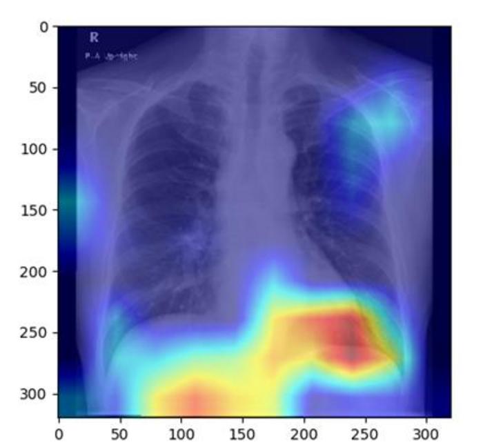
In contrast, chest X-rays represent a more accessible diagnostic tool. These imaging studies are frequently utilized to evaluate the lungs and heart but simultaneously capture portions of the liver. This creates an opportunity for alternative diagnostic approaches for fatty liver disease, an area that has been largely underexplored until now. The synergistic potential of leveraging chest X-ray data to assess liver conditions underscores a critical evolution in medical practice, driven by innovative technology.
The research group utilized a retrospective approach, analyzing a total of 6,599 chest X-ray images derived from 4,414 patients. A noteworthy aspect of their methodology involved the application of controlled attenuation parameter (CAP) scores, which provide quantitative measurements of liver fat content. By training their AI model on this extensive dataset, the researchers were able to enhance its diagnostic accuracy substantially. The area under the receiver operating characteristic curve (AUC) for their model ranged impressively between 0.82 and 0.83, indicating a high level of reliability in distinguishing affected individuals from those without the condition.
Professor Uchida-Kobayashi expressed enthusiasm regarding the implications of their findings. She noted that the introduction of diagnostic methods utilizing readily available chest X-rays suggests the potential for widespread improvements in fatty liver disease detection. The future of such technology could lead to earlier interventions, ultimately altering the disease trajectory for many patients worldwide. Given the current global burden of fatty liver disease, this approach could be transformative in varying healthcare contexts.
The AI-driven detection model represents a convergence of art and science, melding the extensive capabilities of deep learning algorithms with the foundational principles of medical imaging. By enhancing diagnostic pathways, such innovations could streamline patient outcomes limiting the risk of severe disease progression. Such technology resonates with ongoing efforts to integrate artificial intelligence into various healthcare sectors, advocating for smarter, data-driven decision-making processes.
The remarkable implications of this study not only extend to improving diagnostic practices but resonate with larger shifts within the healthcare system. As healthcare professionals navigate the dual challenges of accessibility and accuracy, the AI model provides a beacon of hope for enhancing the standard of care. In particular, hospitals and clinics equipped with basic X-ray technology can bolster their diagnostic capabilities without incurring the substantial costs typically associated with ultrasound or MRI procedures.
Moreover, the researchers’ commitment to verifying their findings through rigorous standards signifies an important step in the journey from innovative research to clinical application. Validation in clinical settings remains paramount for adoption in routine practice, as it helps establish trust and efficacy among healthcare providers and patients alike. As these models continue to evolve, the ethical considerations surrounding AI in healthcare are equally pertinent, calling for responsible integration and transparency.
Furthermore, the initiative encourages a broader dialogue surrounding public health strategies, emphasizing the proactive management of liver health through accessible diagnostic tools. As the global healthcare landscape continues to evolve and respond to increasing rates of liver disease, innovative solutions like the AI model from Osaka Metropolitan University are invaluable. Such endeavors align seamlessly with the vision of empowering healthcare systems worldwide to incorporate cutting-edge technologies that cater to diverse patient needs.
This moment in healthcare reflects not only a technological advancement but also a cultural shift towards embracing innovation for improved health outcomes. By leveraging existing resources—like chest X-rays—this research exemplifies the capability of modern science to reimagine traditional practices in a way that is both efficient and effective. The future landscape of disease diagnosis may very well pivot on the marriage of artificial intelligence with established imaging techniques, heralding a new era in medical diagnostics.
As we witness increased interest in artificial intelligence applications in medicine, this research stands out as a noteworthy example of how AI can serve to bridge gaps in healthcare delivery. Researchers and practitioners alike may draw inspiration from this study, as it highlights the persistent necessity for innovation in health diagnostics, particularly for conditions that carry significant disease burdens like fatty liver disease.
In conclusion, the advent of AI-driven diagnostic tools capable of analyzing chest X-rays for fatty liver disease detection marks a pivotal moment in medical research and clinical practice. This transformative approach aligns seamlessly with the push for more accessible, accurate, and timely diagnoses, ultimately aiming to safeguard patient health and improve quality of life for millions. These advancements exemplify how science continually strives toward solutions that bridge the gaps between technology, medicine, and public health.
Subject of Research: Fatty liver disease
Article Title: Performance of a Chest Radiograph-based Deep Learning Model for Detecting Hepatis Steatosis
News Publication Date: 20-Jun-2025
Web References: Link to the Journal
References: None
Image Credits: Credit: Osaka Metropolitan University
Keywords
AI, Fatty Liver Disease, Chest X-rays, Deep Learning, Imaging Analysis, Medical Research, Healthcare Innovation, Osaka Metropolitan University.
Tags: accessibility of diagnostic toolsadvancements in disease diagnosis technologyAI-driven medical imagingartificial intelligence in healthcarechest X-ray analysis for liver conditionsearly detection of fatty liver diseasefatty liver disease prevalence statisticsimplications of undiagnosed fatty liver diseasemedical imaging and liver healthnon-invasive liver disease detection methodsresearch on AI in medical diagnosticstraditional vs modern diagnostic techniques