
Scientists from the Perelman School of Medicine at the University of Pennsylvania and their colleagues have identified a cell surface receptor protein called TIE2 as a critical component of two key signaling pathways that drive the growth of a type of blood vessel abnormality called cerebral cavernous malformations. In a study, published this week in the Journal of Experimental Medicine, they suggest that drugs targeting TIE2 could prevent CCMs from forming. The findings in mice are detailed in a paper titled “TIE2 links MEKK3–KLF2/4 and PI3K signaling in cerebral cavernous malformation.”
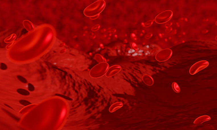
Cerebral cavernous malformations or CCMs are mulberry-shaped vascular lesions that arise in the veins and venules of the central nervous system, resulting in fragile blood vessels with abnormally thin walls. Typically, they are caused by mutations in one of three genes that may be inherited within families but can also arise spontaneously. Once detected, the only way to remove CCMs is by surgical resection but, in many cases, their location within the brain may render them inoperable. If left untreated, these abnormalities can cause brain hemorrhages, strokes, and seizures.
According to the paper, CCM-causing mutations hyperactivate a signaling pathway called the MEKK2-KLF2/4 pathway, in the endothelial cells that line blood vessel walls. This hyperactivation stimulates a second signaling pathway involving the enzyme phosphoinositide 3-kinase (PI3K). Drugs that inhibit the PI3K pathway can prevent the formation of CCMs in mice. But because of the importance of this pathway in many tissues, these drugs can have severe side effects and may be poorly tolerated in the long run.
“Determining how endothelial cells augment PI3K signaling downstream of the MEKK3-KLF2/4 pathway could identify a more blood vessel–specific therapeutic strategy for chronic suppression of CCM growth,” according to Mark Kahn, MD, a professor at Penn, and senior author of the new study. “However, the molecular mechanism by which augmented MEKK3-KLF2/4 function increases PI3K signaling has remained unclear.”
In this new study, Kahn and his colleagues reveal that these two key signaling pathways are linked by TIE2, a receptor protein on the surface of endothelial cells that regulates blood vessel development. Specifically, they report that TIE2 activity was enhanced in the endothelial cells surrounding both human and mouse CCMs. Levels of the TIE2 protein increased in response to elevated MEKK3-KLF2/4 signaling, which increased the activation of the PI3K pathway. Crucially, they showed that inhibiting TIE2 with rebastinib, an orally bioavailable small-molecule inhibitor of multiple tyrosine kinases, prevented the development of new CCMs in mice.
Overall, the data suggests that “pharmacologic blockade of TIE2 may provide an endothelial cell-centered approach for chronic suppression of CCM disease with fewer side effects than systemic PI3K pathway inhibition,” Kahn said.