
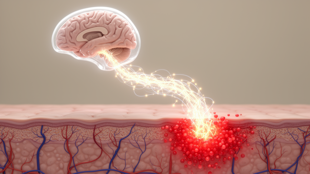
A new investigation reveals that the primary somatosensory cortex actively amplifies cutaneous inflammation in animal models of rosacea and psoriasis rather than functioning merely as a passive recipient of peripheral sensations. Rosacea is widely recognized as a chronic inflammatory dermatosis characterized by recurrent facial flushing, persistent erythema, telangiectasia, papules, pustules, and debilitating sensory symptoms such as burning, stinging, and heightened sensitivity to environmental and thermal changes. While traditional dermatological frameworks have primarily focused on localized cutaneous neuroimmune dysregulation and abnormal vascular reactivity in lesional tissue, the study demonstrates that defined higher-order cerebral circuits in the primary somatosensory cortex, designated as S1, causally modulate the magnitude of inflammatory responses in the skin through pathways dependent on peripheral sensory nerves.
To investigate the anatomical and functional pathways connecting peripheral skin tissue with central sensory structures, investigators initiated retrograde viral tracing studies in wild-type female C57BL/6J mice housed under specific pathogen-free conditions. Researchers administered cutaneous injections of pseudorabies virus expressing enhanced green fluorescent protein, designated PRV-EGFP, into the dorsal skin of mice to identify retrogradely labeled neuronal populations in coronal cerebral sections. This skin-directed viral tracing successfully labeled distinct cortical neurons located in both the primary somatosensory cortex and the secondary somatosensory cortex, confirming direct multi-trans-synaptic neural connectivity between peripheral skin sites and higher-order cortical regions. The researchers subsequently administered noxious cutaneous stimuli, including intradermal formalin and topical capsaicin applications, to determine whether these identified circuits become activated during acute tissue distress.
Analysis of the immediate early gene c-Fos demonstrated robust neuronal recruitment within S1 following noxious sensory stimulation. Immunofluorescence examinations showed a substantial overlap between retrogradely labeled PRV-positive neurons and c-Fos-expressing cells in the somatosensory cortex. Beyond acute chemical stimulation, the investigators sought to evaluate whether established models of chronic cutaneous inflammation similarly engage this cortical hub. In mice subjected to daily intradermal injections of LL37 antimicrobial peptide at a concentration of 320 micromolar to induce rosacea-like dermatitis, S1 c-Fos expression increased significantly. An identical elevation in cortical c-Fos induction occurred in mice exposed to daily topical applications of five percent imiquimod cream on the right ear over six consecutive days to induce psoriasiform dermatitis, establishing that diverse forms of cutaneous inflammation engage somatosensory cortical networks.
The study also examined the involvement of primary somatosensory cortex architecture in human patients through neuroimaging. Investigators recruited 102 rosacea patients and 79 age- and sex-matched healthy control volunteers, acquiring resting-state functional magnetic resonance imaging scans on a Siemens Prisma 3T system. All patients underwent rigorous clinical assessments, including the Clinician Erythema Assessment, Investigator Global Assessment, flushing evaluations, telangiectasia scoring, burning symptom severity, and quality-of-life measurements. Analysis focused on low-frequency fluctuation amplitude, abbreviated as ALFF, and regional homogeneity, abbreviated as ReHo, within the bilateral postcentral gyrus, the human anatomical homologue of S1. While initial univariate comparisons between the entire rosacea cohort and healthy controls showed no statistically significant differences in left or right S1 metrics, exploratory threshold-based subgroup analyses revealed that patients bearing higher clinical and psychological burdens, such as elevated flushing, anxiety, depression, and stress, exhibited lower spontaneous activity and reduced regional homogeneity in the right S1.
To establish whether S1 activation plays an active causal role in driving skin pathology, the team deployed chemogenetic tools via stereotaxic cranial surgery in mice. Adeno-associated viral vectors encoding either excitatory hM3Dq or inhibitory hM4Di designer receptors exclusively activated by designer drugs were precisely injected into S1 coordinates. Systemic administration of clozapine-N-oxide, delivered intraperitoneally at two milligrams per kilogram daily starting two days prior to inflammation induction, allowed selective modulation of S1 neuronal firing. Conversely, chemogenetic inhibition of S1 significantly attenuated every measured pathological parameter.
Molecular and cellular evaluations confirmed that cortical activation translated into robust immunological alterations within lesional skin. Quantitative real-time polymerase chain reaction assays revealed that chemogenetic excitation of S1 markedly elevated transcript levels of pro-inflammatory cytokines, specifically tumor necrosis factor, interleukin-1 beta, and interleukin-6, alongside elevations in interleukin-1 alpha and interleukin-36 gamma. Immunofluorescence microscopy further demonstrated extensive dermal recruitment and accumulation of CD4-positive T lymphocytes in rosacea lesions following S1 stimulation, whereas chemogenetic silencing of S1 significantly diminished both cytokine expression and CD4-positive cell infiltration. Importantly, when investigators manipulated S1 activity in uninjured skin injected only with phosphate-buffered saline, neither chemogenetic activation nor inhibition produced erythema, epidermal thickening, cytokine upregulation, or leukocyte infiltration, demonstrating that S1 does not trigger de novo disease but acts as an amplifier of ongoing peripheral inflammation.
Global quantitative liquid chromatography-tandem mass spectrometry skin proteomics provided comprehensive mechanistic insight into the downstream biological cascades regulated by S1 activity. Differential expression analysis in skin tissue resolved 342 differentially expressed proteins when comparing S1-activated mice to controls, and 513 differentially expressed proteins when comparing S1-inhibited mice to controls. Trend analyses fitted across the inhibition, control, and activation axis identified graded protein alterations directly tracking cortical activity. Gene set enrichment analysis against Hallmark pathways established that S1 activation drove pronounced enrichment of pro-inflammatory and pro-vascular programs, including coagulation cascades, angiogenesis, complement activation, interferon responses, and interleukin-6-JAK-STAT3 and tumor necrosis factor-NF-kappa-B signaling cascades, alongside negative enrichment of oxidative phosphorylation and mTORC1 signaling. Biological process gene ontology terms supported extensive vascular transport, leukocyte migration, and sensory-neural remodeling, corroborated by CD31 staining showing increased vascular density under S1 activation and reduced density under S1 inhibition.
The study sought to determine whether this central amplificatory loop relies on peripheral sensory innervation. The authors administered resiniferatoxin, a potent TRPV1 receptor ligand that selectively ablates and desensitizes peripheral sensory afferents, via subcutaneous abdominal injections at escalating doses of 30, 70, and 100 micrograms per kilogram in four-week-old mice. Following sensory denervation, the severe aggravation of rosacea-like dermatitis normally triggered by chemogenetic S1 activation was largely eliminated, resulting in significantly smaller erythematous areas, reduced redness scores, diminished skin swelling, decreased histological infiltration, and reduced cytokine levels. Furthermore, sensory ablation dramatically suppressed the induction of activating transcription factor 3, known as ATF3, within primary sensory neurons of the dorsal root ganglia. In contrast, sympathetic denervation achieved through systemic 6-hydroxydopamine administration failed to prevent S1-mediated aggravation in dermatitis models, indicating that descending central influence is mediated predominantly via sensory rather than sympathetic fibers.
Similar regulatory mechanisms operated in imiquimod-induced psoriasiform dermatitis, where chemogenetic S1 excitation markedly exacerbated ear swelling, scaling, erythema, epidermal hyperplasia, Ki67 proliferation markers, and Gr-1-positive neutrophil and CD4-positive T-cell infiltration. Consistent with the rosacea model, chemogenetic inhibition or resiniferatoxin-mediated sensory ablation significantly reduced psoriasiform severity and suppressed dorsal root ganglion ATF3 induction. These convergent findings indicate that the primary somatosensory cortex functions as a broad cortical amplifier across multiple sensory-linked inflammatory skin disorders, maintaining a bidirectional dialogue wherein peripheral inflammation recruits cortical sensory circuits, which in turn feed back onto peripheral sensory nerves to intensify local tissue pathology.
Capitalizing on these physiological mechanisms, the researchers tested a non-invasive therapeutic intervention using repetitive transcranial magnetic stimulation targeted directly to the S1 region. Mice were treated with continuous theta-burst stimulation, an inhibitory magnetic stimulation protocol delivering 600 pulses per session consisting of three-pulse bursts at 50 Hertz repeated at 5 Hertz, administered once daily for five days prior to model induction and twice daily thereafter. Cortical inhibitory stimulation produced substantial therapeutic improvements in both rosacea-like and psoriasiform mouse models, demonstrating visible reductions in lesion thickness, marked decreases in clinical erythema scores, suppressed tissue cytokine expression, and reduced immune-cell infiltration. By establishing that non-invasive cortical neuromodulation can interrupt neuroimmune disease progression, these findings introduce the primary somatosensory cortex as an accessible central target for future clinical therapies aimed at treating refractory neuroinflammatory skin conditions.
Subject of Research: Neural regulation of rosacea-like skin inflammation by the primary somatosensory cortex
Article Title: Primary somatosensory cortex activation aggravates rosacea-like dermatitis in a sensory nerve-dependent manner
Article References: Zhang, Y., Li, T., Yan, Q., Xiao, X., Duan, Y., Li, Y., Zhao, S., Chen, M., Deng, Z., Wang, B., Huang, Y., Tang, J., Hu, X., Liu, L., Zhang, Z., Deng, X., Xie, H., Zhao, H., & Li, J. (2026). Primary somatosensory cortex activation aggravates rosacea-like dermatitis in a sensory nerve-dependent manner. Biomarker Research, 14(1), Article 163. https://doi.org/10.1186/s40364-026-01043-6
Image Credits: AI Generated
DOI: 10.1186/s40364-026-01043-6
Keywords: primary somatosensory cortex, rosacea, neuroimmune regulation, sensory nerves, dermatitis, chemogenetics, c-Fos, resting-state fMRI, repetitive transcranial magnetic stimulation, resiniferatoxin, LL37 peptide, psoriasiform dermatitis, dorsal root ganglia, ATF3, cutaneous inflammation, somatosensory circuits, brain-skin axis

