
In a groundbreaking leap for synthetic biology and cellular engineering, a team of researchers spearheaded by Professor Hiroaki Suzuki at Chuo University has unveiled a pioneering method for fabricating artificial cells embedded with DNA condensates that mimic the natural nuclei of eukaryotic cells. This breakthrough capitalizes on advanced microfluidic technologies to mass-produce highly uniform lipid bilayer vesicles—also known as liposomes—each containing intricately structured DNA condensates serving as model nuclei. By replicating these hierarchical DNA arrangements inside artificial cells, the scientists are pushing the frontier closer to creating synthetic cell systems capable of complex biological functions akin to living organisms.
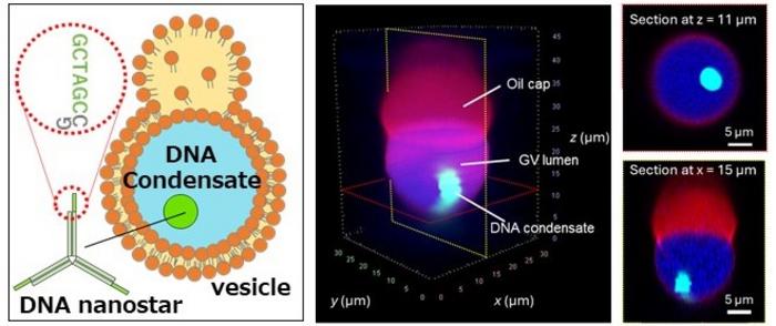
The core of this innovation lies in the precise harnessing of microfluidic devices, which serve as production lines capable of generating monodisperse giant unilamellar vesicles with remarkable reproducibility. These vesicles encapsulate key components such as DNA nanostars and ionic salts essential for the formation of condensed DNA structures. Unlike traditional methods that often rely on thermal annealing—a process that can damage sensitive biomolecules—this technique utilizes osmotic pressure to modulate vesicle volume and thereby concentrate molecular constituents within the artificial cells. This elegant volume-control mechanism facilitates the spontaneous assembly of controlled DNA condensates without compromising the integrity of other enzymatic or protein synthesis systems encapsulated within the vesicles.
DNA nanostars, the building blocks used in this system, are unique DNA nanostructures composed of multiple single-stranded DNA arms branching in Y-shaped or X-shaped configurations. These arms are engineered with short, complementary “sticky ends” that guide precise hybridization and network formation. By fine-tuning these interactions, the researchers can program the DNA condensates to exhibit distinct physical properties and maintain a fine balance between condensation and dissociation dynamics. This controlled assembly recapitulates aspects of natural chromatin compaction found in cell nuclei—a feat that holds immense promise for bottom-up construction of artificial cellular compartments.
.adsslot_1ZsKhBTFpy{ width:728px !important; height:90px !important; }
@media (max-width:1199px) { .adsslot_1ZsKhBTFpy{ width:468px !important; height:60px !important; } }
@media (max-width:767px) { .adsslot_1ZsKhBTFpy{ width:320px !important; height:50px !important; } }
ADVERTISEMENT
One of the most compelling outcomes of this study is the successful demonstration of protein synthesis directly from genes housed within the model nuclei. Embedded inside these synthetic vesicles, the genes encoding green fluorescent protein (GFP) were transcribed and translated, evidencing that the artificial nuclei serve not merely as inert DNA storage, but as functional hubs capable of molecular information processing. This crucial achievement paves the way for future artificial cells to transcend static biomimicry and actively engage in biological activities such as gene expression, signal transduction, and environmental responsiveness.
Recent trends in synthetic cell research have increasingly focused on reproducing the complex spatial and functional hierarchies observed in living eukaryotic cells. Central to this challenge is the recreation of the nucleus—not only the largest organelle but the epicenter of genetic regulation. By innovating a scalable and reproducible method for assembling artificial nuclei within uniform vesicles, the research team has established a versatile platform that rigorously controls the physicochemical environments within these synthetic cells. This precision engineering is critical for faithfully emulating the nuanced behavior of intracellular organelles and holds potential for sophisticated applications ranging from biosensing to therapeutic cell replacement.
The strategic use of microfluidic devices confers several advantages over conventional liposome preparation techniques. Traditional methods generally produce vesicles with wide size distributions and inconsistent encapsulation efficiencies, limiting the fidelity of artificial cell models. Here, the microfluidic approach exploits fluid dynamics in micron-sized channels to consistently form vesicles with near-identical diameters and uniform molecular content. Such homogeneity is essential not only for reliable experimental reproducibility but also for future industrial-scale manufacturing of artificial cell systems for biomedical use.
Moreover, circumventing the need for thermal annealing during DNA condensate formation is a significant technical advancement. Thermal cycles employed in prior approaches can denature proteins or destabilize fragile enzymatic complexes integral to synthetic cell functions. By orchestrating DNA nanostar condensation via osmotic volume adjustments, the researchers preserve the functionality of sensitive biomolecules encapsulated within the artificial cells. This gentle yet effective methodology broadens the spectrum of molecules that can coexist within synthetic cell constructs, unlocking multifaceted biomimetic capabilities.
These artificial cells—with their model nuclei capable of genetic activity—open exciting new frontiers in the creation of artificial biological systems tailored for specific tasks. For instance, molecular recognition functions imbued by DNA nanostars can enable these synthetic entities to detect and respond to environmental cues. The ability to program artificial cells with such responsiveness may revolutionize targeted drug delivery, biosensing, and regenerative medicine, where cellular substitutes need to integrate seamlessly within living tissues or dynamically interact with their surroundings.
In addition to their synthetic biology significance, the study carries profound implications for understanding the fundamental principles governing intracellular organization. By reconstructing DNA condensation processes in vitro within confined lipid membranes, the team offers a novel experimental platform to dissect physical and chemical factors underlying chromatin packing and gene regulation. Insights gleaned from these artificial nuclei might thus inform not only technological innovation but also basic biological research into cell nucleus structure-function relationships.
The research has been rigorously validated through experimental studies, culminating in the publication of comprehensive results in the high-impact journal JACS Au. The team’s multidisciplinary collaboration intertwines expertise from materials science, molecular biology, and microfluidics engineering. Such confluence of disciplines exemplifies the integrative efforts required to tackle the immense complexity of synthetic cell construction and highlights the potential for continued cross-field partnerships in advancing artificial life sciences.
Looking forward, this study establishes a foundational step toward realizing fully functional artificial cells that could one day replace or augment natural cells in therapeutic contexts. The scalability and reproducibility afforded by the microfluidic-based assembly line model position this technology well for future translational research and commercial development. As artificial cells evolve to perform increasingly sophisticated biological processes, they may revolutionize the approaches to disease treatment, personalized medicine, and environmental biosensing.
In summary, the controlled formation of DNA condensates as model nuclei within monodisperse giant vesicles represents a milestone achievement in synthetic cell engineering. By fusing the precision of microfluidics with the programmability of DNA nanotechnology, the research team brings us closer to the dream of building artificial life forms from the bottom up. This breakthrough not only enriches our understanding of cellular organization but also unlocks transformative possibilities for biomedical technologies that harness the power of synthetic biology.
Subject of Research: Cells
Article Title: Controlled Formation of DNA Condensates as Model Nuclei in Monodisperse Giant Vesicles
News Publication Date: 18-Jun-2025
Web References:
https://pubs.acs.org/journal/jaaucr
http://dx.doi.org/10.1021/jacsau.5c00568
References:
Ushiyama, R., Koiwai, K., Suzuki, H. (2021). Plug-and-play microfluidic production of monodisperse giant unilamellar vesicles using droplet transfer across water-oil interface. Sensors and Actuators B: Chemical, 355, 131281.
Ushiyama, R., Nanjo, S., Tsugane, M., Sato, R., Matsuura, T., Suzuki, H. (2024). Identifying condition for protein synthesis inside giant vesicles using microfluidics toward standardized artificial cell production. ACS Synthetic Biology, 13(1), 68-76.
Image Credits: Chuo University
Keywords: Artificial cells, DNA condensates, microfluidics, DNA nanostars, lipid bilayer vesicles, synthetic biology, protein synthesis, osmotic control, model nuclei, bottom-up cell construction, GFP expression
Tags: advanced cellular engineering methodsartificial cell mass productioncontrolled DNA condensation techniquesDNA condensates in artificial cellseukaryotic cell mimickinghierarchical DNA arrangement replicationlipid bilayer vesicles fabricationmicrofluidic technology in synthetic biologymodel nuclei in synthetic cell systemsmonodisperse giant unilamellar vesiclesosmotic pressure in cell engineeringsynthetic biology breakthroughs