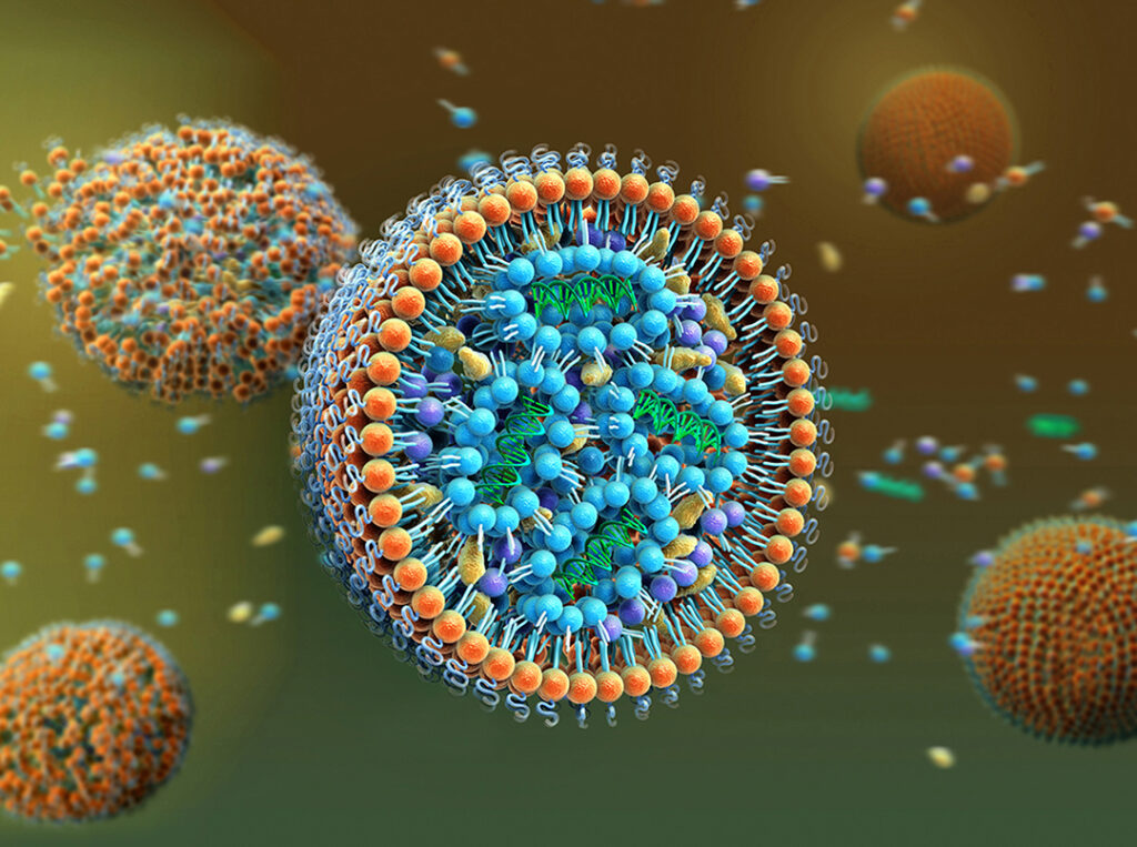
Using cryogenic mass spectrometry, scientists from the University of Nottingham’s School of Pharmacy and their collaborators elsewhere developed an approach for depth profiling frozen lipid nanoparticles (LNPs) to reveal the layers and orientation of the constituent molecules. Their research provides insights into the relative positions of each component within lipid nanoparticles that could help clarify LNP behavior and contribute to the design of new formulations used in RNA vaccine and drug delivery.
Details of the work are published in a Communications Chemistry paper titled, “Study on Molecular orientation and stratification in RNA-lipid nanoparticles by Cryogenic Orbitrap Secondary Ion Mass Spectrometry.”
The success of the Moderna and Pfizer BioNTech COVID-19 vaccines showcased the value of LNPs for delivering RNA-based therapies. Besides vaccines, LNPs are also used to deliver other types of treatments, including small-interfering RNAs for rare hereditary diseases, and they could also be used in therapies for various cancers, pulmonary conditions, and more.
Understanding how components on the surface of LNPs behave is essential for making them more efficient and safe for use in therapies. As the researchers wrote, “the identity of the lipids that present at their surface play a role in how they interact with and are perceived by the body and their resultant potency.” Furthermore, insights into the surface structure could also help with quality control during scale-up of biomanufacturing processes from the laboratory to clinical applications.
Commenting on the study, Robert Langer, PhD, one of the co-authors on the paper and an institute professor in the department of chemical engineering at Massachusetts Institute of Technology, noted that while “effective drug delivery relies on an intricate mix of molecules in lipid nanoparticles to effectively deliver RNA therapeutics” they can have varying efficacies and be difficult to engineer. “This research provides a new way of characterizing and understanding the make-up of lipid nanoparticles, which could pave the way for engineering more potent and targeted LNPs to enable the broadest application of RNA therapies for all types of diseases,” he said.
The research team, which also included scientists from Sail Biomedicines and the U.K.’s National Physical Laboratory, utilized Cryogenic Orbitrap secondary ion microscopy (Cryo-OrbiSIMS) to obtain structural details of the lipid nanoparticles. This high-pressure freezing cryo-preparation facility keeps biological samples maintained close to their native state.
“This cryogenic molecular surface and interfacial analysis advance” makes it possible to characterize “the native surface of delicate hydrated pharmaceutical systems used in the body” something that has been a significant challenge for some time, noted Morgan Alexander, PhD, a professor of biomedical surfaces at University of Nottingham’s School of Pharmacy and lead author on the study. “We expect to apply this new method to many systems, including lipid nanoparticles, other pharmaceutical delivery systems, and hydrated biomaterials.”
Kerry Benenato, PhD, chief platform officer at Sail Biomedicines, added that “by enabling precise surface characterization, the technology that the team has developed paves the way for the engineering by design of LNP-based medicines with tunable properties, including biodistribution, thereby expanding the potential of RNA-based therapeutics.”