A single number extracted from a routine MRI scan before treatment appears to predict how long patients with recurrent IDH-mutant gliomas will live without tumor progression after receiving bevacizumab, according to a new retrospective study from researchers at the University of California, Los Angeles. The finding, published in the Journal of Neuro-Oncology, extends a biomarker concept originally developed for glioblastoma into a slower-growing but still dangerous family of brain tumors, and it may offer clinicians a practical tool for deciding which recurrent glioma patients are most likely to benefit from an expensive antiangiogenic drug that helps some patients far more than others.
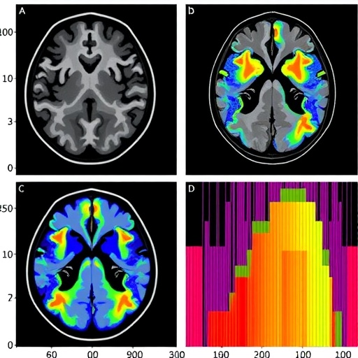
The biomarker in question is called ADC-L, the mean of the lower Gaussian peak in a histogram of apparent diffusion coefficient (ADC) values measured within contrast-enhancing tumor tissue. ADC is derived from diffusion-weighted MRI and quantifies the random Brownian motion of water molecules in tissue. Water diffuses more freely in sparse, loosely packed tissue and more slowly in densely cellular tissue, which is why ADC is widely used as a noninvasive proxy for tumor cellularity. In the two-compartment histogram model used by the UCLA team, the ADC values of every voxel inside the enhancing tumor volume are fitted with a double Gaussian mixture, mathematically expressed as a weighted sum of a lower distribution and a higher distribution, each with its own mean and standard deviation. The lower peak, ADC-L, is thought to represent the most cellular, most restricted fraction of the enhancing tumor, and it is this parameter that has repeatedly stratified survival in recurrent IDH-wildtype glioblastoma patients treated with anti-VEGF agents such as bevacizumab.
Whether the same principle held for IDH-mutant gliomas was far from obvious. IDH mutations in the isocitrate dehydrogenase enzymes define a biologically distinct subtype of adult glioma, present in more than 80 percent of lower-grade WHO grade 2 and 3 gliomas and most common in adults under 50. These tumors produce the oncometabolite 2-hydroxyglutarate, which drives CpG island hypermethylator phenotype (CIMP) and frequent MGMT promoter methylation, rendering them unusually sensitive to alkylating chemotherapy and radiation. They also tend to have lower cellularity and intrinsically higher mean ADC values than IDH-wildtype glioblastoma, raising the possibility that the ADC-L signal could be diluted or masked. Moreover, the phase II TAVAREC trial had found no progression-free or overall survival benefit from adding bevacizumab to temozolomide in recurrent WHO grade 2/3 gliomas without 1p/19q codeletion, casting doubt on the value of antiangiogenic therapy in this population and underscoring the need for a biomarker that could enrich patient selection.
To test the hypothesis, the researchers assembled a cohort of sixty patients with IDH-mutant recurrent glioma, comprising 41 astrocytomas and 19 oligodendrogliomas, all treated with bevacizumab at UCLA between 1998 and 2024. From an eligible pool of 97 patients, 21 were excluded for lacking pretreatment diffusion-weighted imaging and 16 for insufficiently sized enhancing tumors, under one milliliter, or for image distortion that precluded reliable ADC analysis. Every patient had measurable contrast-enhancing disease. The contrast-enhancing tumor volumes were segmented automatically using the NS-HGlio artificial intelligence segmentation tool, with manual quality control by trained lab members under the supervision of an experienced neuroradiologist. ADC values were then extracted from all voxels within the enhancing volume and fitted with the double Gaussian model using nonlinear regression, with the mean of the lower Gaussian component, ADC-L, designated the primary imaging biomarker.
The analysis produced a strikingly clean answer. The optimal ADC-L threshold for risk stratification, determined by scanning all possible cutoffs and calculating Mantel-Haenszel hazard ratios for each, was 1.19 square micrometers per millisecond, remarkably close to the 1.2 to 1.24 micrometers squared per millisecond thresholds previously established for recurrent IDH-wildtype glioblastoma across more than seven trials and over 400 patients. Patients whose tumors exhibited ADC-L at or above this value had a median progression-free survival of 5.46 months, compared with just 2.76 months for those below the threshold, a statistically significant difference with a hazard ratio of 2.3 in univariate analysis. Intriguingly, despite the clear separation in progression-free survival, no difference in overall survival was observed between the two groups, a point the authors interpret in the context of the many subsequent therapies available to IDH-mutant patients after progression, which muddy the interpretability of overall survival as an endpoint.
Multivariable Cox regression, adjusting for the number of prior recurrences, confirmed that low ADC-L was an independent predictor of shorter progression-free survival, with a hazard ratio of 2.054 and a p-value of 0.0228. Astrocytoma histology, as opposed to oligodendroglioma, was also independently predictive, with a hazard ratio of 2.295 and a p-value of 0.0158. When the analyses were stratified by histopathological subtype, ADC-L remained a significant predictor within both groups: in astrocytoma patients, low ADC-L carried a hazard ratio of 2.1, and in oligodendroglioma patients the effect was even larger in magnitude, with a hazard ratio of 5.5, though the small oligodendroglioma subgroup of only 19 patients limits confidence in that estimate. The similarity of the optimal threshold across IDH genotypes is perhaps the most intriguing technical finding, suggesting a conserved biological mechanism linking restricted water diffusion within enhancing tumor to anti-VEGF responsiveness regardless of the underlying mutation status.
The authors offer a speculative but biologically plausible explanation for the diffusion-based stratification. All patients in the cohort had previously undergone chemoradiation, and increased diffusivity within enhancing tumor tissue after cytotoxic therapy is well documented in gliomas. Elevated diffusivity after radiation has also been linked to a higher likelihood of post-treatment reactive changes such as pseudoprogression. It is conceivable, the authors suggest, that ADC-L effectively captures the tumor’s reaction to prior therapy, with higher diffusivity reflecting a less densely viable, less aggressively proliferative enhancing volume that is primed for a better response when vascular endothelial growth factor signaling is subsequently blocked. This interpretation would reconcile the biomarker’s apparent transferability across two diseases with quite different baseline cellularity and microstructure.
The study also leveraged modern response assessment methodology, which strengthens its findings. Progression was scored according to RANO 2.0 criteria without confirmation scans, using volumetric measurements of contrast-enhancing tumor from three-dimensional segmentations processed by an automated RANO calculator, with progression defined as either at least 40 percent volumetric enlargement of the enhancing tumor or unequivocal non-enhancing progression on T2-weighted images, the latter evaluated by a neuroradiologist blinded to the diffusion data. In a landmark analysis anchored to the time of RANO determination, the combination of high pretreatment ADC-L and achievement of a radiographic response was strongly associated with improved progression-free survival, with a p-value of 0.00034. Percent reduction in enhancing tumor volume and confirmed RANO response were also significant univariate predictors, reinforcing the consistency of the overall picture.
The limitations of the work are acknowledged candidly by the investigators. The retrospective, single-center design spanned 26 years, during which MRI scanner hardware, diffusion sequences, and treatment protocols evolved considerably, and ADC measurements were not harmonized across scanners, potentially introducing variability that attenuated statistical associations. The total cohort of 60 patients is modest, the oligodendroglioma subgroup is underpowered, and because the 1.19 micrometers squared per millisecond threshold was derived and tested within the same cohort, overfitting cannot be excluded, making external validation in an independent IDH-mutant cohort essential before prospective clinical use. The cohort was also restricted to patients with measurable contrast-enhancing disease, likely a higher-grade recurrence phenotype enriched for malignant transformation, so the findings may not extend to tumors that progress primarily through T2/FLAIR expansion without enhancement. Finally, because contrast enhancement reflects blood-brain barrier disruption rather than tumor per se, and no patients underwent re-resection at the time of bevacizumab initiation, the fraction of the enhancing volume representing viable tumor versus treatment-related change cannot be determined with certainty.
Even with those caveats, the implications are meaningful. Bevacizumab, a humanized monoclonal antibody targeting VEGF, is widely used in recurrent high-grade gliomas but delivers inconsistent benefit, and the TAVAREC trial’s negative result has left clinicians with little guidance on which IDH-mutant patients, if any, should receive it. The present cohort, treated later in the disease course with bevacizumab as the primary salvage agent, may represent a biologically distinct population enriched for angiogenic dependence, and pretreatment ADC-L combined with enhancing tumor burden may be precisely the tool needed to identify that subgroup. The authors argue that progression-free survival, which isolates the effect of the treatment under investigation without the confounding influence of post-progression therapies, is the more relevant endpoint for this disease, and they call for prospective evaluation of ADC-L as a biomarker for bevacizumab patient selection in IDH-mutant recurrent glioma. If validated externally, the biomarker could be deployed almost immediately in clinical practice, since diffusion-weighted imaging is already part of standard brain tumor MRI protocols and the histogram analysis can be computed from existing scans without additional cost or radiation exposure.
Subject of Research: Pretreatment ADC histogram analysis (ADC-L) as a predictive imaging biomarker of progression-free survival in recurrent IDH-mutant gliomas treated with bevacizumab
Subject of Research: Cancer
Article Title: Diffusion histogram analysis predicts progression-free survival in contrast enhancing recurrent IDH mutant gliomas treated with bevacizumab
Article References: Loxterkamp, E., Luo, A., Sanvito, F., Le, C. T., Raymond, C., Yang, C., Prins, T. J., Fisher, A., Salamon, N., Liau, L. M., Chong, R. A., Nghiemphu, P. L., Nathanson, D. A., Cloughesy, T. F., Lai, A., & Ellingson, B. M. (2026). Diffusion histogram analysis predicts progression-free survival in contrast enhancing recurrent IDH mutant gliomas treated with bevacizumab. Journal of Neuro-Oncology, 179(2), Article 75. https://doi.org/10.1007/s11060-026-05782-2
Image Credits: AI Generated
DOI: 10.1007/s11060-026-05782-2
Keywords: IDH-mutant glioma, ADC-L, diffusion histogram analysis, bevacizumab, progression-free survival, predictive biomarker, apparent diffusion coefficient, anti-VEGF therapy, astrocytoma, oligodendroglioma, MRI biomarker, recurrent glioma
Cite Scienmag News
APA MLA Chicago
Nathaniel Bowman. (September 10, 2026). Diffusion histogram analysis forecasts survival in bevacizumab-treated recurrent IDH-mutant gliomas. Scienmag. https://scienmag.com/diffusion-histogram-analysis-forecasts-survival-in-bevacizumab-treated-recurrent-idh-mutant-gliomas/
Copy citation Download RIS
Tags: ADC-L biomarker in recurrent gliomasADC-L prognostic indicator in brain tumorsapparent diffusion coefficient in tumor imagingbevacizumab treatment response in IDH-mutant gliomasbiomarkers for glioma prognosisdiffusion histogram analysisdiffusion MRI biomarker for recurrent gliomasdiffusion-weighted MRI in brain tumor managementdiffusion-weighted MRI in neuro-oncologyglioma survival predictionhistogram analysis of apparent diffusion coefficienthistograms of ADC values for survival forecastingMRI-based prognostic tools for brain tumorsMRI-based tumor cellularity measurementneuro-oncology imaging techniquesnoninvasive glioma prognosis toolnoninvasive tumor cellularity assessmentpersonalized treatment planning for recurrent gliomaspersonalized treatment planning in recurrent gliomaspredictive imaging biomarkers for brain tumor therapytumor cellularity and treatment outcomes