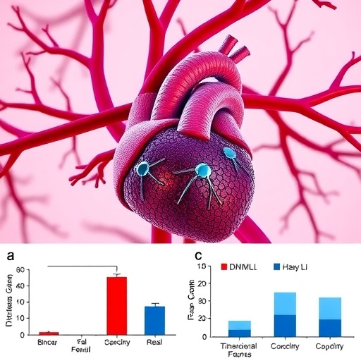
In a groundbreaking study published in Pediatric Research in early 2025, researchers have uncovered pivotal insights into the mechanisms by which mutations in the DNM1L gene contribute to cardiac dysfunction through mitochondrial impairment. Leveraging the transformative potential of human induced pluripotent stem (iPS) cell technology, this research pushes the boundaries of our understanding of mitochondrial dynamics and their critical role in heart health. The study holds promise not only for elucidating the pathogenesis of hereditary cardiac diseases but also for steering future therapeutic strategies that target mitochondrial function at the cellular level.
Mitochondria, often referred to as the powerhouses of the cell, are essential organelles that regulate energy production, calcium homeostasis, and apoptosis. Their dynamic nature, characterized by continuous fission and fusion, is crucial for maintaining cellular health and metabolic adaptability. The DNM1L gene encodes dynamin-related protein 1 (Drp1), a key regulator of mitochondrial fission. Mutations in this gene disrupt the delicate balance, causing abnormal mitochondrial morphology and consequent bioenergetic deficits. The research team, led by Osawa, Fujita, and Kagami, employed patient-derived iPS cells to model these mutations in vitro, overcoming previous limitations of animal models and providing a human-specific mechanistic perspective.
By differentiating iPS cells into cardiomyocytes, the study recreated a disease-relevant cellular environment, unveiling how defective Drp1 impacts mitochondrial architecture and function within heart cells. The mutated Drp1 compromised mitochondrial division, leading to elongated and dysfunctional mitochondria. This morphological aberration correlated with a decrease in mitochondrial membrane potential and ATP synthesis, impairing the cardiomyocytes’ ability to meet the high energetic demands essential for proper cardiac contractility.
The team’s meticulous analysis extended to mitochondrial respiratory chain function, revealing significant reductions in the activities of complexes I and IV. These complexes are integral components of oxidative phosphorylation, driving the production of ATP via electron transport. Impaired complex function culminated in diminished oxygen consumption rates and elevated reactive oxygen species (ROS) generation, reinforcing the link between DNM1L mutations and oxidative stress-induced cellular damage. Excess ROS may exacerbate mitochondrial dysfunction, creating a deleterious feedback loop that accelerates cardiomyocyte injury and contributes to the progression of cardiac disease.
Electrophysiological assessments of the iPS-derived cardiomyocytes revealed altered calcium handling dynamics, which are critical for synchronized heart contractions. The mutated cells exhibited irregular calcium transients and delayed cytosolic calcium clearance, suggesting that mitochondrial dynamics influence calcium cycling and, by extension, cardiac rhythm stability. This novel connection highlights potential mechanisms underlying arrhythmias observed in patients harboring DNM1L mutations.
Importantly, the study also explored the activation status of mitophagy—the selective autophagic removal of damaged mitochondria. The defective Drp1-mediated fission impaired the segregation of dysfunctional mitochondria, hindering their clearance and resulting in the buildup of defective organelles. Accumulated mitochondrial damage likely precipitates cellular stress responses and contributes to cardiomyocyte apoptosis, both key features of cardiac remodeling and dysfunction.
From a therapeutic standpoint, the findings pave the way for interventions aimed at restoring mitochondrial dynamics. Experiments applying molecular agents that modulate fission and fusion proteins showed partial recovery of mitochondrial morphology and function, emphasizing the potential reversibility of these defects. This revelation fuels hope for precision medicine approaches tailored to ameliorate or prevent cardiac complications linked to DNM1L mutations.
Moreover, this research underscores the versatility and power of human iPS cell technology as a platform for modeling rare genetic heart diseases. Unlike conventional animal studies, patient-derived iPS cells encapsulate the precise genetic context, allowing for personalized pathophysiological investigations and drug screenings. The study thereby sets a precedent for integrating stem cell biology with mitochondrial medicine to transform disease modeling and therapeutic development.
The work also contributes significantly to our understanding of mitochondrial biology in the cardiac setting. While mitochondrial dysfunction has long been implicated in a range of cardiomyopathies, dissecting the specific molecular derangements caused by DNM1L mutations affords clarity in the intricate network of mitochondrial quality control processes. It challenges existing paradigms and suggests that targeted modulation of mitochondrial dynamics could become an innovative cornerstone of cardiac therapeutics.
This research arrives at a particularly critical juncture in cardiovascular medicine, where heart failure and cardiomyopathies remain leading causes of morbidity and mortality worldwide. Patient populations with genetic mitochondrial defects currently face limited treatment options. The mechanistic insight into DNM1L mutations’ role in cardiac mitochondrial impairment offers a beacon of potential for developing novel treatment avenues that transcend symptom management to address disease etiology.
Furthermore, the elevation of ROS and subsequent oxidative stress identified in this study may link DNM1L-related cardiac dysfunction to broader metabolic and inflammatory pathways. This intersection presents fertile ground for future studies aiming to unravel the systemic consequences of mitochondrial anomalies and their interplay with cardiovascular health.
In conclusion, the Osawa et al. study embodies a significant advance in cardiac mitochondrial pathology research. By exploiting the unique capabilities of human iPS cell-based models, it elucidates how DNM1L mutations disrupt mitochondrial fission and function, culminating in profound cardiac cellular impairment. As the scientific community continues to probe the complexities of mitochondrial dynamics, this work stands out as a compelling demonstration of how gene-specific defects translate into disease phenotypes and offers tangible prospects for targeted therapies.
Moving forward, comprehensive investigations are warranted to determine if these cellular phenotypes manifest similarly in vivo and to evaluate the efficacy of proposed therapeutic compounds in clinical settings. The ripple effects of this research also invite exploration into other tissues affected by DNM1L mutations, potentially broadening the impact of these findings beyond the heart.
These discoveries underscore the transformative power of integrating genetic, cellular, and molecular techniques in decoding complex diseases. The convergence of cutting-edge stem cell technology with mitochondrial biology heralds a new era of precision cardiovascular medicine, providing hope for patients suffering from devastating mitochondrial cardiac disorders.
Subject of Research: Cardiac dysfunction caused by mitochondrial impairment due to DNM1L mutations assessed using human induced pluripotent stem (iPS) cells.
Article Title: Cardiac dysfunction due to mitochondrial impairment assessed by human iPS cells caused by DNM1L mutations.
Article References:
Osawa, M.T., Fujita, Y., Kagami, K. et al. Cardiac dysfunction due to mitochondrial impairment assessed by human iPS cells caused by DNM1L mutations. Pediatr Res (2025). https://doi.org/10.1038/s41390-025-04045-6
Image Credits: AI Generated
DOI: https://doi.org/10.1038/s41390-025-04045-6
Tags: bioenergetic deficits in heart diseasecardiac dysfunction mechanismsDNM1L gene mutationsDrp1 role in mitochondrial fissionhereditary cardiac disease pathogenesishuman-induced pluripotent stem cellsiPS cell technology in researchmitochondrial dynamics in cardiomyocytesmitochondrial impairment in heart cellsmitochondrial morphology and cellular healthpatient-derived iPS cell modelstherapeutic strategies targeting mitochondria


