
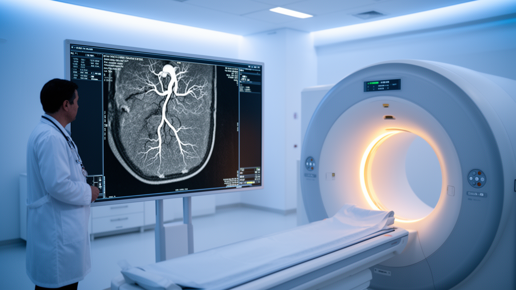
Radiologists in China have shown that a carefully tuned dual-energy CT protocol can image the inferior vena cava — the body’s largest vein — with roughly one-fifth less contrast medium, one-third less iodine, and a modestly lower radiation dose than a conventional 120 kVp scan, while producing image quality and reader confidence that were statistically indistinguishable from the standard approach. The single-center prospective study, conducted at Suining Central Hospital and published in Frontiers in Cardiovascular Medicine, combined 60 keV virtual monoenergetic imaging with a 60% strength of an iterative reconstruction algorithm called ASIR-V, and its authors present the optimized combination as a practical option for routine venous imaging.
The inferior vena cava returns approximately two-thirds of the venous blood from the abdominopelvic organs and the legs to the right atrium, and it is the highway along which a surprising range of pathology travels or takes root. Congenital variants such as a duplicated or left-sided cava, acquired problems such as tumor invasion, Budd-Chiari syndrome, and thrombosis, and the presence of implanted filters or stents all demand clear depiction of the vessel lumen. CT angiography has become a preferred noninvasive tool for this job, but the conventional way of doing it carries a familiar trade-off: high-concentration iodinated contrast, typically 350 to 400 mgI/mL injected at relatively large volumes, is needed to make the venous lumen stand out, and that iodine load raises concern about contrast-induced acute kidney injury, especially in patients with impaired renal function or those who need repeated examinations.
Dual-energy CT offers a way out of that bind. The scanner used in the study, a 256-row Revolution CT system, rapidly switches its X-ray tube between 80 and 140 kVp within a single rotation, acquiring near-simultaneous low- and high-energy data. From that paired acquisition the machine can synthesize 101 virtual monoenergetic image datasets spanning 40 to 140 keV, each simulating how the tissue would look if imaged with X-rays of a single energy. Lower energies, near the iodine K-edge of 33.2 keV, dramatically amplify iodine attenuation and vascular contrast, but the photons penetrate less tissue, fewer of them reach the detectors, and quantum noise rises. Higher energies clean up noise and beam-hardening artifacts but wash out contrast. Choosing the right energy is therefore a balancing act, and it is one that previous venous imaging studies had addressed only partially.
The team enrolled 150 consecutive patients scheduled for inferior vena cava CT angiography between January and December 2025 and, after excluding 30 for reasons such as diffuse stenosis, severe occlusion, motion artifacts, or non-diagnostic image quality, analyzed 120. Allocation was sequential rather than randomized: patients scanned from January through June formed the conventional Group A, and those scanned from July through December formed the dual-energy Group B, with 60 patients in each. The groups were well matched in age, sex, height, weight, body mass index, and the mix of IVC findings, which included thrombosis, stenosis, filter placement, and normal vessels. Group A received the standard recipe — 120 kVp scanning with iomeprol at 400 mgI/mL delivered at 1.5 mL per kilogram of body weight, reconstructed with 50% ASIR-V. Group B received gemstone spectral imaging with a leaner contrast strategy — iodixanol at 320 mgI/mL at 1.2 mL per kilogram.
The optimization proceeded in two steps. First, with ASIR-V held at 50%, seven virtual monoenergetic datasets from 40 to 70 keV in 5 keV increments were reconstructed for every Group B patient. Objective measurements — CT attenuation, standard deviation within the vessel, background noise in adjacent muscle, signal-to-noise ratio, and contrast-to-noise ratio — were taken at five venous levels from the hepatic IVC down to the femoral veins. The physical trend was unambiguous: as energy increased, attenuation, noise, SNR, and CNR all fell progressively. The 40 keV images boasted the highest attenuation, SNR, and CNR, but also the greatest noise and, crucially, the worst subjective scores. Two blinded radiologists rating image quality on a 5-point Likert scale gave 40 keV averages of roughly 3.1, versus 4.8 at 60 keV, which scored highest and differed significantly from every other energy level. Raw signal metrics alone, the authors caution, would have pointed to the wrong choice.
The second step fixed the energy at 60 keV and varied reconstruction strength. Images were rebuilt with filtered back projection — the classical, noise-prone algorithm — and with ASIR-V at 20%, 40%, 60%, 80%, and 100%. As expected, rising iterative strength steadily suppressed vascular noise and background noise while inflating SNR and CNR: at 100% ASIR-V the hepatic IVC SNR reached a median of 28.48, more than four times the filtered-back-projection value of 6.16. But the readers did not celebrate. Scores climbed from FBP through 60% ASIR-V and then declined, because the strongest reconstructions produced an over-smoothed, plastic-like texture that radiologists find unnatural and distrust. Sixty percent ASIR-V, which cut hepatic IVC noise to a median of 14.80 HU while preserving texture, won on both objective balance and subjective preference — a middle path consistent with prior reports in coronary, pediatric, and chest CT.
With the winning combination defined as Group B1, the head-to-head comparison followed. The optimized dual-energy images actually showed higher IVC attenuation, higher vascular standard deviation, and higher background noise than the conventional 120 kVp images, all statistically significant. Yet the ratios that matter most to interpretation — SNR and CNR — did not differ significantly between protocols, and mean subjective image-quality scores were nearly identical at 4.75 for the conventional group and 4.80 for the optimized group, with strong inter-observer agreement. Diagnostic-confidence ratings for thrombosis, luminal stenosis, filter position, and normal IVC findings were likewise similar, and no thrombus was missed and no filter misclassified in either group. The authors are careful to note that these confidence scores measure reader interpretability, not diagnostic accuracy against a reference standard.
The dose savings were substantial and consistent. Median effective dose fell from 10.07 mSv in the conventional group to 8.81 mSv in the dual-energy group, a reduction of about 12.51%. Median contrast volume dropped from 93.75 mL to 75.00 mL, roughly 20%, and median iodine intake fell from 37.50 g to 24.00 g, a 36% cut — all differences significant at P < 0.001. Because the leaner contrast strategy pairs a lower concentration with a lower volume per kilogram, the protocol may be particularly attractive for patients who need serial imaging, such as filter or thrombosis follow-up, or for those in whom contrast must be managed cautiously. The authors stop short of any renal claim: serum creatinine and estimated glomerular filtration rate were not measured before or after scanning, so the iodine reduction cannot yet be linked to a lower incidence of contrast-induced kidney injury.
The study’s limitations frame how far its conclusions should travel. It was a non-randomized, single-center feasibility study of modest size, optimization and evaluation were performed in the same cohort, and no reference standard or formal equivalence design was used — so the absence of significant differences should not be read as proof of equivalence. The optimal energy was selected at one reconstruction strength and the optimal strength at that energy, without a full-factorial exploration of interactions. All data came from one vendor’s platform, limiting generalizability, and only ASIR-V was tested; newer deep-learning reconstruction methods remain unexamined. The subgroup analyses of diagnostic confidence were exploratory and not powered to detect differences, and the authors label the whole optimized protocol hypothesis-generating, calling for multicenter randomized trials with appropriate reference standards.
Even with those caveats, the message is a concrete one for imaging departments. Rather than optimizing a single dial, the study stitched together three levers — reduced contrast concentration and volume, an evidence-based monoenergetic energy, and a moderate iterative reconstruction strength — and showed that the combination held image quality steady while trimming iodine and radiation exposure. Within the 40 to 70 keV range and the FBP-to-100% reconstruction range evaluated, 60 keV with 60% ASIR-V emerged as the sweet spot for venous imaging of the inferior vena cava, a result that, if validated more broadly, could make lower-dose IVC angiography a realistic default rather than an experimental luxury.
Subject of Research: Optimization of a dual-energy CT protocol for inferior vena cava CT angiography using virtual monoenergetic imaging and iterative reconstruction
Article Title: Optimized dual-energy CT protocol for inferior vena cava CT angiography using 60 keV virtual monoenergetic imaging and 60% ASIR-V
Article References: Deng, J., Fan, X., Zhang, Q., Yan, J., Li, Y., Yan, G., Zhao, L., Shen, X., Peng, Q., Ma, T., Wu, S., Wang, X., HE, X., McClure, M. A., & Bhetuwal, A. (2026). Optimized dual-energy CT protocol for inferior vena cava CT angiography using 60 keV virtual monoenergetic imaging and 60% ASIR-V. Frontiers in Cardiovascular Medicine, 13, Article 1969018. https://doi.org/10.3389/fcvm.2026.1969018
Image Credits: AI Generated
DOI: 10.3389/fcvm.2026.1969018
Keywords: dual-energy CT, inferior vena cava, CT angiography, virtual monoenergetic imaging, ASIR-V, iodinated contrast, radiation dose, image quality, iterative reconstruction, venous thrombosis, IVC filter, contrast-induced kidney injury