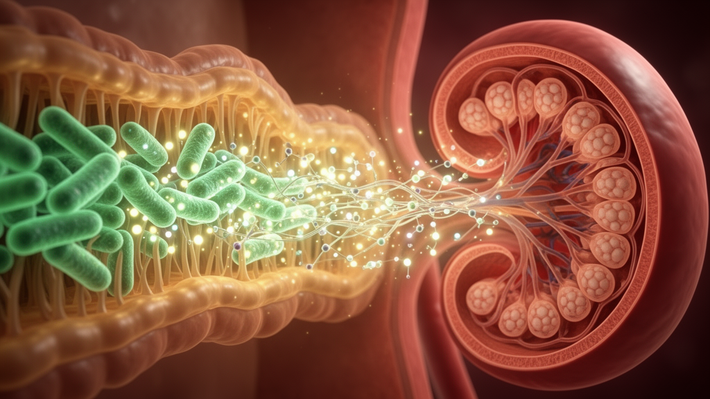
A narrative review published in Frontiers in Cell and Developmental Biology outlines how disruptions in the gut microbiome and imbalances in microbial metabolites drive the onset and progression of diabetic kidney disease. The synthesis, led by researchers Yuan Yuan, XinXin Pang, Yachan Gao, and colleagues from Henan University of Chinese Medicine, evaluates evidence across 132 selected references following a search of PubMed, Web of Science, and Embase through August 2026. Diabetic kidney disease represents a major microvascular complication of diabetes mellitus and constitutes a leading global driver of end-stage renal disease. The authors trace the functional crosstalk along the gut-kidney axis, demonstrating that a shift from beneficial fermentation to toxic accumulation establishes a self-reinforcing pathogenic loop that damages renal tissue and accelerates functional decline.
The review details the pathophysiological foundation of diabetic kidney disease, which begins with persistent hyperglycemia and diabetic tubulopathy that persists throughout the course of diabetes. Within China, a nationwide cross-sectional study cited by the authors indicated that the weighted prevalence of chronic kidney disease among individuals with diabetes reached 32.6 percent. Sustained high glucose activates pro-inflammatory cascades, prompting the release of cytokines including tumor necrosis factor-alpha and interleukin-1 beta, while shedding endothelial glycocalyx and downregulating tight-junction proteins to compromise the glomerular filtration barrier. Concurrently, activation of the polyol, protein kinase C, and hexosamine pathways promotes the accumulation of advanced glycation end products, which bind their receptor to generate excessive reactive oxygen species. Hyperglycemia also activates the renin-angiotensin-aldosterone system, induces autophagic impairment, and provokes lipid metabolic dysfunction that triggers apoptosis, ferroptosis, and stress-induced senescence in renal tubular epithelial cells.
As renal filtration deteriorates, the intestinal environment undergoes marked taxonomic and functional shifts, transitioning from protective saccharolytic fermentation to detrimental proteolytic fermentation. At the phylum level, systematic evidence encompassing 578 patients with diabetic kidney disease and 444 healthy controls confirmed a distinct expansion of Gram-negative Proteobacteria alongside Actinobacteria and Bacteroidetes, accompanied by a significant contraction of Firmicutes. This expansion directly elevates the gut-derived load of lipopolysaccharide endotoxins. Analysis of overall microbial diversity revealed marked decreases in richness, Chao, Ace, and Shannon indices, alongside an elevated Simpson index. These diversity metrics demonstrate clinical correlations: while Chao, Ace, and Shannon indices correlate positively with estimated glomerular filtration rates and act as protective indicators against disease severity, the Simpson index operates as an independent risk factor for worsening renal injury.
At the genus level, shotgun metagenomic and 16S ribosomal RNA sequencing demonstrate a bidirectional dysbiosis characterized by the depletion of beneficial short-chain fatty acid producers and the proliferation of opportunistic pathogens. Genera including Faecalibacterium, Roseburia, Bifidobacterium, Phocaeicola, and Lachnospira undergo widespread decline. Conversely, pathogenic taxa such as Escherichia, Shigella, Enterocloster, Klebsiella, Enterococcus, Mediterraneibacter, and Fusobacterium become enriched. Linear discriminant analysis effect size confirmed Shigella and Enterocloster as signature biomarkers of diabetic kidney disease. Depletion of health-associated taxa correlates negatively with clinical markers of injury including glycated hemoglobin, blood urea nitrogen, and the urinary albumin-to-creatinine ratio, while pathogen expansion correlates with elevated circulating pro-inflammatory mediators including interleukin-6, soluble CD14, and an altered CD4-to-CD8 T cell ratio.
The loss of short-chain fatty acid producers severely compromises systemic and renal homeostasis by diminishing protective levels of acetate, propionate, and butyrate. Under normal conditions, these microbial fermentation products engage G-protein coupled receptors, specifically GPR43 and GPR109A, to suppress the nuclear factor-kappa B inflammatory axis and activate nuclear factor erythroid 2-related factor 2 antioxidant pathways. Animal models cited in the review confirm that high-fiber diets or direct fatty acid supplementation attenuate renal damage in diabetic mice, an effect almost completely abolished in GPR43- or GPR109A-knockout strains. Mechanistically, GPR43 activation enhances binding between beta-arrestin-2 and I-kappa-B-alpha to prevent phosphorylation of the p65 subunit. Beyond direct signaling, butyrate fuels colonic epithelial cells, stimulates zonula occludens-1 expression to preserve the intestinal mechanical barrier, and limits bacterial endotoxin translocation. Propionate and butyrate also upregulate renal organic anion transporter 1, promoting the secretory clearance of circulating uremic toxins.
In contrast to the loss of protective molecules, the accumulation of gut-derived uremic toxins exacerbates renal parenchymal destruction. Indoxyl sulfate and p-cresyl sulfate represent two major protein-bound toxins arising from microbial degradation of dietary tryptophan and tyrosine or phenylalanine, followed by hepatic sulfation. Due to extensive protein binding, both molecules exhibit poor clearance via conventional dialysis and accumulate as tubular secretion declines. These toxins trigger nicotinamide adenine dinucleotide phosphate oxidase activity, driving excessive reactive oxygen species generation, activating cyclic adenosine monophosphate response element-binding protein signaling, and inducing transforming growth factor-beta 1 and tissue inhibitor of metalloproteinases 1 to accelerate epithelial-mesenchymal transition and interstitial fibrosis.
Circulating concentrations of this metabolite rise sharply in diabetic kidney disease due to enhanced microbial production combined with impaired glomerular filtration and tubular secretion. Clinical cohorts demonstrate that serum levels correlate positively with urinary albumin-to-creatinine ratios and negatively with estimated glomerular filtration rates, serving as an independent risk factor for disease progression. In diabetic rat models, fecal microbiota transplantation experiments verified that donor microbiota possess enhanced capacity for metabolite synthesis, driving heightened Smad3 phosphorylation and renal fibrosis. High-glucose environments further stimulate SUMO2/3-mediated post-translational modification of Smad4, which amplifies pro-fibrotic signaling, suppresses peroxisome proliferator-activated receptor alpha and gamma expression, inhibits fatty acid oxidation, and prompts intracellular lipid deposition.
Bile acid metabolism represents another pivotal pathway disrupted by intestinal dysbiosis along the gut-kidney axis. Primary bile acids synthesized from hepatic cholesterol undergo bacterial transformation mediated by bile salt hydrolase and 7-alpha-dehydroxylase enzymes. In diabetic kidney disease, dysbiosis alters the ratios of conjugated to unconjugated and primary to secondary bile acids. Total serum bile acid levels dropping below 2.8 millimoles per liter independently predict progression toward end-stage renal disease. Renal proximal tubular cells exhibit marked downregulation of the apical sodium-dependent bile acid transporter and the farnesoid X receptor, which prevents reabsorption of taurocholic acid and suppresses protective signaling. Conversely, cytotoxic conjugated species such as glycochenodeoxycholic acid accumulate in the plasma, where their concentrations correlate inversely with glomerular filtration rate, directly driving oxidative stress, tubular apoptosis, and interstitial matrix accumulation.
These mechanistic insights support several targeted therapeutic strategies designed to restore microbial equilibrium and mitigate renal decline. High-fiber dietary interventions selectively enrich butyrate-producing bacteria, suppressing pro-inflammatory and pro-fibrotic transcription in tubular cells and podocytes. Very low-protein diets supplemented with essential amino acid analogues demonstrate measurable reductions in serum indoxyl sulfate, while Mediterranean dietary patterns lower circulating levels of uremic toxins. Supplementation with probiotics, including Bifidobacterium and Lactobacillus strains, alongside prebiotics such as inulin and resistant starches, reduces serum p-cresyl sulfate and replenishes fecal short-chain fatty acids in chronic kidney disease cohorts. Experimental fecal microbiota transplantation from healthy donors similarly reconstructed microbial diversity, restored cholesterol homeostasis, and attenuated glomerulosclerosis and tubular damage in diabetic animal models, while natural products like berberine suppressed toxin synthesis and replenished beneficial commensals.
Despite encouraging findings across preclinical investigations, the authors caution that substantial methodological and translational limitations remain. The current evidentiary base relies heavily on rodent experiments, in vitro assays, and small clinical cohorts that predominantly employed 16S ribosomal RNA profiling rather than high-resolution metagenomics. Crucial clinical confounders—including varied antibiotic regimens, background diets, glycemic status, and concurrent antihypertensive medications—were rarely controlled or standardized across original reports. Moreover, rigorous randomized controlled trials evaluating hard renal outcomes, such as sustained reductions in filtration rate or rates of kidney failure, remain lacking for dietary regimens, probiotic strains, and microbial transplantation. Establishing the clinical validity of gut-kidney axis therapies will require prospective multicenter longitudinal studies, standardized multi-omics pipelines, and robust causal inference frameworks to confirm therapeutic efficacy in human populations.
Subject of Research: Pathogenic mechanisms and therapeutic targeting of the gut-kidney axis in diabetic kidney disease
Article Title: Gut microbiota and its metabolites in diabetic kidney disease: underlying mechanisms and recent research breakthroughs
Article References: Yuan, Y., Pang, X., Gao, Y., Wang, Y., Du, C., Qiao, Z., & Cheng, R. (2026). Gut microbiota and its metabolites in diabetic kidney disease: underlying mechanisms and recent research breakthroughs. Frontiers in Cell and Developmental Biology, 14, 2007740. https://doi.org/10.3389/fcell.2026.2007740
Image Credits: AI Generated
DOI: 10.3389/fcell.2026.2007740
Keywords: diabetic kidney disease, gut microbiota, gut-kidney axis, short-chain fatty acids, butyrate, indoxyl sulfate, p-cresyl sulfate, trimethylamine N-oxide, bile acids, dysbiosis, Proteobacteria, Firmicutes, renal fibrosis, uremic toxins, TLR4/NF-κB pathway, NLRP3 inflammasome, fecal microbiota transplantation