
Liver cancer stands as one of the most prevalent forms of cancer worldwide, ranking as the sixth most common type and, crucially, one of the leading causes of cancer-related mortality. For clinicians and medical practitioners, the accurate segmentation of liver tumors within imaging scans is a fundamental aspect of effective disease management. However, despite its importance, manual segmentation tasks performed by radiologists can be remarkably time-consuming and often introduce significant variability that is dependent on the radiologist’s level of experience and expertise. In a groundbreaking shift toward overcoming these challenges, advances in artificial intelligence (AI) are proving transformative, particularly through the development of AI-based tumor segmentation models.
Traditional deep learning approaches, particularly convolutional neural networks, have ushered in a new era of tumor assessment in medical imaging. These AI systems are adept at identifying and delineating the contours of tumors from medical scans, thereby offering a degree of precision that can greatly enhance diagnostic accuracy. However, the effectiveness of these models is traditionally linked to large datasets, often requiring thousands of cases—typically anywhere between 1,000 to 10,000 unique examples—to train effectively. This requirement for extensive data presents a significant hurdle, particularly in specialized medical fields where access to large datasets may be markedly limited.
In a striking development, researchers at the Institute of Science Tokyo, led by Professor Kenji Suzuki and PhD candidate Yuqiao Yang, have pioneered a novel AI model designed to segment liver tumors from computed tomography (CT) scans. This model operates effectively even with exceedingly small training datasets, exceeding the performance benchmarks set by existing state-of-the-art systems. This significant advancement was documented in a recent publication in the esteemed journal IEEE Access, published on May 16, 2025. The introduction of this innovative approach is set to make waves in the realm of AI-driven medical imaging and may reshape existing paradigms of research and diagnosis.
.adsslot_nU7iaMCJS8{ width:728px !important; height:90px !important; }
@media (max-width:1199px) { .adsslot_nU7iaMCJS8{ width:468px !important; height:60px !important; } }
@media (max-width:767px) { .adsslot_nU7iaMCJS8{ width:320px !important; height:50px !important; } }
ADVERTISEMENT
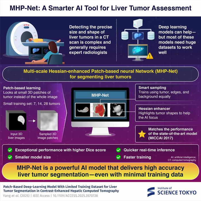
Central to this innovation is the multi-scale Hessian-enhanced patch-based neural network, more succinctly referred to as MHP-Net. This cutting-edge architecture is designed to dissect complex medical images into smaller, manageable 3D patches. By focusing on one segment of the image at a time, the model can achieve better accuracy in segmentation. Each of these small patches is then paired with an enhanced version derived through a sophisticated technique known as Hessian filtering, which accentuates spherical objects—such as tumors—within the image. This meticulous process enables MHP-Net to generate high-resolution tumor segmentation maps that effectively distinguish liver tumors from contrast-enhanced CT scans.
To validate the model’s performances, the research team employed the “Dice similarity score,” a widely recognized metric for assessing the congruence between the predicted segmentation and the actual annotated images created by expert radiologists. The Dice score ranges from 0 to 1, with closer values to 1 indicating higher accuracy. Remarkably, even with limited training sets comprising just 7, 14, and 28 tumor examples, MHP-Net achieved impressive Dice scores of 0.691, 0.709, and 0.719, respectively. These results indicate that the model not only performs well but also surpasses that of many established models such as U-Net, Res U-Net, and HDense-U-Net.
Beyond its promising performance metrics, the lightweight design of MHP-Net affords rapid training times of fewer than 10 minutes, alongside real-time inference that operates within approximately four seconds per patient. This rapid processing capability underscores the model’s potential suitability for implementation in clinical environments that often operate under resource constraints.
Professor Suzuki articulates the transformative potential of this research: “This is just a start in the field of small-data AI, where meaningful and clinically relevant deep learning models can be built from limited datasets.” He further alludes to MHP-Net’s broader implications, suggesting that its successful architecture could serve as a template for developing small-data AI solutions across various sectors of medical imaging, including the detection of rare cancers.
The study underscores a pivotal moment in AI-driven medical analysis, shedding light on the practical applications of small-data methodologies. The ability to democratize access to advanced AI technologies by requiring fewer data facilitates the integration of these tools into under-resourced healthcare settings, where access to comprehensive datasets is often a significant barrier. As the framework established by MHP-Net evolves, researchers are keen to explore its scalability and effectiveness across diverse clinical applications, championing the potential for cost-effective and versatile AI deployments in healthcare globally.
Furthermore, the researchers envision extending the application of MHP-Net beyond liver tumor segmentation, propelling the model toward addressing a wide array of medical imaging challenges. This ambition not only highlights the versatility of AI but also the potential it holds to revolutionize healthcare delivery and patient outcomes in regions where resources and data might be scarce.
This study illuminates a stimulating frontier in artificial intelligence’s role within medicine. The pioneers at the Institute of Science Tokyo are setting in motion significant shifts that will impact how medical imaging techniques are employed in diagnosis and treatment planning. MHP-Net embodies the aspirations of researchers to leverage technology for a greater social good, fostering innovation that resonates deeply within the foundations of medical science.
In summary, as liver cancer continues to prevail as a significant global health challenge, developments such as those pioneered by the researchers at the Institute of Science Tokyo offer glimmers of hope. This technological leap not only marks a significant stride toward redefining tumor segmentation practices but also catalyzes the broader potential for AI to reshape the landscape of medical imaging. MHP-Net stands as a testament to what is achievable when innovation meets necessity, presenting a compelling narrative about the future of healthcare.
Subject of Research:
Article Title: Patch-Based Deep-Learning Model With Limited Training Dataset for Liver Tumor Segmentation in Contrast-Enhanced Hepatic Computed Tomography
News Publication Date: 16-May-2025
Web References: http://dx.doi.org/10.1109/ACCESS.2025.3570728
References: IEEE Access, Volume 13
Image Credits: Institute of Science Tokyo, Japan
Keywords
Artificial intelligence, Medical treatments, Tomography, Liver tumors, Medical imaging, Liver cancer, Biomedical engineering, Medical technology
Tags: accuracy in liver cancer detectionadvanced imaging techniques in oncologyAI-based liver tumor segmentationartificial intelligence in healthcareconvolutional neural networks in medical imagingenhancing cancer treatment through technologyimproving diagnostic precision with AIlarge datasets in deep learning modelsliver cancer mortality and diagnosismanual vs automated tumor segmentationovercoming challenges in cancer diagnosistransformative AI applications in radiology