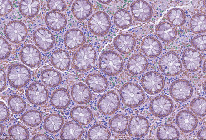

Histopathological data remain one of the most trusted tools in science when doctors or researchers want to understand what’s happening inside a tissue. Today, they have largely gone digital. These images, therefore, contain enormous information about tissues from different scales.
However, these images remain separate from modern multimodal and single-cell frameworks. While genetics and single-cell biology have developed effective ways for sharing and comparing data, digital pathology images are hard to incorporate—stored in proprietary formats, processed with incompatible tools, and hard to connect to molecular information like RNA sequencing. Thus, the valuable resources of digitalized tissue images are largely underutilized in many research and clinical settings.
Now, a new study introduces LazySlide, an open-source Python package built on “the scverse ecosystem for efficient whole-slide image analysis and multimodal integration” designed to make whole-slide image analysis more accessible, interoperable, and ready to plug into the same computational workflows that already drive modern genomics. Through the power of foundation models, LazySlide aims to democratize digital pathology analysis by bridging histopathology with omics workflows.
“Histology contains an enormous amount of biological information, but it is often difficult to access computationally,” says Yimin Zheng, PhD, a postdoc in the lab of André Rendeiro, PhD, at the CeMM Research Center for Molecular Medicine in Vienna, Austria. “With LazySlide, we wanted to provide a tool that allows researchers to explore tissue images in a systematic, quantitative way and to connect what they see under the microscope with underlying molecular processes.”
The study, published in Nature Methods in the paper, “LazySlide: accessible and interoperable whole slide image analysis” highlights how flexible tools like LazySlide can accelerate research and help bridge the gap between tissue structure and molecular function—an essential step toward a more integrated understanding of health and disease.
LazySlide enables scientists to break down whole-slide images into smaller, manageable regions and analyze them using advanced AI models that can recognize patterns in tissue structure, identify different cell types, and quantify subtle changes in tissue architecture, without requiring extensive manual annotation.
The study shows that visual information from tissue images can be directly linked to molecular data such as gene expression profiles. In one example, the researchers analyzed artery tissue samples with and without calcification. LazySlide not only distinguished healthy from diseased tissue based on image features alone, but also revealed biological pathways, such as inflammatory signaling, that became visible only when image data and RNA sequencing data were analyzed together.
By using AI models that link visual patterns to text concepts, researchers can ask questions such as where signs of calcification appear within a tissue sample. The software then highlights relevant regions and generates quantitative scores, turning visual impressions into measurable data.
This approach also allows “zero-shot” analysis: LazySlide can recognize the organ of origin of tissue samples or distinguish healthy from diseased tissue without being specifically trained for each task. This greatly lowers the barrier for applying advanced image analysis methods in biomedical research.
LazySlide was designed to integrate seamlessly with existing computational biology tools widely used in genomics and single-cell research. By making digital pathology more interoperable with these established workflows, the software helps bring tissue imaging into the broader ecosystem of data-driven life sciences.
“Our goal was to make whole-slide image analysis more accessible and more connected to the kinds of data researchers already use every day,” says Rendeiro. “By treating tissue images as rich datasets rather than static pictures, we can gain new insights into how diseases shape human biology.”