
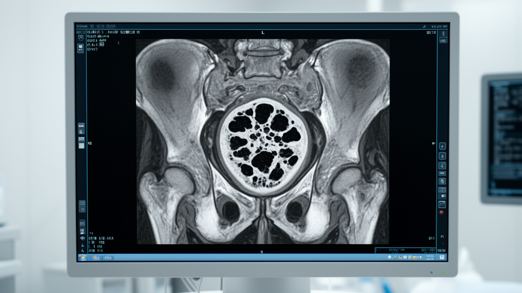
A systematic review and meta-analysis published in the Journal of Orthopaedic Surgery and Research shows that greater magnetic resonance imaging-derived necrotic burden is strongly associated with subsequent femoral head collapse in patients with osteonecrosis of the femoral head. Femoral head collapse substantially influences the clinical management of osteonecrosis of the femoral head. Although magnetic resonance imaging can quantify the spatial extent of necrotic lesions before radiographic collapse takes place, the strength, precision, and generalizability of the association between imaging-derived necrotic burden and subsequent collapse have remained uncertain. To address this uncertainty, an investigative team led by Zixuan Wang, Qingwen Zhang, and colleagues conducted a systematic review evaluating quantitative imaging measures in relation to collapse.
The systematic review protocol was registered in the PROSPERO international prospective register under registration identifier CRD420261456638. The researchers executed comprehensive literature searches across seven major medical and bibliographic databases from their inception dates up to August 8, 2026. The searched databases included PubMed, Embase, the Cochrane Library and CENTRAL, Web of Science, the China National Knowledge Infrastructure, Wanfang Data, and the VIP database. Quantitative magnetic resonance imaging-derived measures identified across the retrieved literature were synthesized separately within three distinct frameworks: a lesion-burden framework, a necrotic-angle framework, and a continuous measurement framework.
Random-effects meta-analyses were conducted using restricted maximum likelihood estimation paired with Knapp-Hartung-adjusted confidence intervals and ninety-five percent prediction intervals. The authors performed additional sensitivity analyses to evaluate the influence of threshold derivation strategies, minimum follow-up duration requirements, temporal sequencing of magnetic resonance imaging assessment before documented collapse, and individual-study influence on the pooled estimates. Methodological quality and risk of bias across the included cohort studies were appraised using the Quality in Prognosis Studies tool. The overall certainty of the synthesized prognostic evidence was evaluated according to the Grading of Recommendations Assessment, Development and Evaluation framework for prognostic factors.
Following study-level eligibility screening and an extraction re-audit, eighteen reports representing sixteen cohort families were included in the quantitative synthesis. In the primary binary analysis pooling six studies involving 383 hips, higher magnetic resonance imaging-quantified necrotic burden was associated with greater odds of subsequent collapse, yielding an odds ratio of 31.62 with a ninety-five percent confidence interval of 11.70 to 85.50. Between-study statistical heterogeneity remained low, with an I-squared value of 23.8 percent. The calculated ninety-five percent prediction interval ranged from 5.60 to 178.67, confirming that an elevated necrotic burden consistently portends increased odds of collapse across comparable clinical settings.
Synthesis of the necrotic-angle metrics showed marked estimation instability despite an elevated average summary odds ratio. Across four studies involving 336 hips that evaluated necrotic-angle parameters, the pooled odds ratio was 52.55, with a ninety-five percent confidence interval of 6.33 to 436.25 and an I-squared heterogeneity statistic of 63.8 percent. However, the ninety-five percent prediction interval was exceptionally wide, spanning from 0.20 to 14,090.93 and crossing the null boundary of one. This wide interval indicates that although necrotic-angle measurements were strongly associated with collapse on average, predictions for future individual cohorts exhibit profound statistical uncertainty.
In the continuous measurement framework, which synthesized data from six studies involving 359 hips, hips that subsequently collapsed exhibited a greater baseline necrotic burden, yielding a pooled Hedges’ g of 2.23 with a ninety-five percent confidence interval of 1.29 to 3.16. Nevertheless, substantial between-study heterogeneity was observed, reflected by an I-squared value of 86.0 percent. The ninety-five percent prediction interval for this continuous analysis spanned from minus 0.31 to 4.76, crossing the null threshold and demonstrating that greater baseline necrotic volume or area cannot be assumed to predict subsequent collapse in every prospective clinical setting.
When the authors restricted their sensitivity analysis strictly to unequivocally pre-collapse imaging assessments across four studies involving 231 hips, the pooled Hedges’ g was 1.83, with a ninety-five percent confidence interval of 0.51 to 3.14 and a ninety-five percent prediction interval spanning from minus 1.82 to 5.48. Although sensitivity analyses generally preserved the positive direction of average associations, several prediction intervals crossed the null boundary. Furthermore, application of the GRADE framework resulted in a low certainty rating for the primary binary analysis and very low certainty ratings for both the necrotic-angle and continuous measurement frameworks.
The investigators concluded that while higher magnetic resonance imaging-derived necrotic burden is associated with subsequent femoral head collapse in osteonecrosis of the femoral head, the true magnitude of this association is imprecisely estimated and varies across measurement approaches and study settings. Imaging-derived volume, area, surface, angular, and spatial measures should be regarded as related but non-interchangeable prognostic markers. Current evidence does not support a universal imaging threshold, a stand-alone treatment rule, or a validated individual-risk prediction model. Prospective multicenter validation using clearly pre-collapse imaging assessments, standardized measurements, predefined thresholds, adequate follow-up duration, and appropriate handling of bilateral hips remains necessary.
Subject of Research: Prognostic association between MRI necrotic burden and femoral head collapse in osteonecrosis
Article Title: MRI-derived necrotic burden and subsequent femoral head collapse in osteonecrosis of the femoral head: a systematic review and meta-analysis
Article References: Wang, Z., Li, Y., Xie, L., Wang, Z., Yang, F., & Zhang, Q. (2026). MRI-derived necrotic burden and subsequent femoral head collapse in osteonecrosis of the femoral head: a systematic review and meta-analysis. Journal of Orthopaedic Surgery and Research. https://doi.org/10.1186/s13018-026-07315-z
Image Credits: AI Generated
DOI: 10.1186/s13018-026-07315-z
Keywords: osteonecrosis of the femoral head, femoral head collapse, magnetic resonance imaging, necrotic burden, systematic review, meta-analysis, prognostic factors, necrotic angle, Hedges’ g, prediction intervals, QUIPS risk of bias, GRADE assessment, PROSPERO registration, pre-collapse MRI, orthopedic surgery, restricted maximum likelihood

