Satiety, the sensation of being full, or sated, is an important neurobiological process for maintaining body weight, but individuals can still have the desire to eat sweet, high-sugar-containing foods, even when feeling full after a meal. Studying mice, and also some human volunteers, a research team led by scientists at the Max Planck Institute for Metabolism Research discovered that a type of neuron that promotes satiety also switches on what the investigators call “sugar appetite,” which then drives overconsumption.
Henning Fenselau, PhD, research group leader at the Max Planck Institute for Metabolism Research, and colleagues, reported their findings in Science, in a paper titled, “Thalamic opioids from POMC satiety neurons switch on sugar appetite.” The team suggests the findings could lead to new research into neural circuits that may be relevant to the development of treatments for obesity.
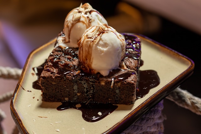
“Satiety is triggered by caloric surplus and in response to the resolution of caloric deficiency, such as after eating a meal,” the authors explained. However, they further pointed out, “Although feeding behavior and overall food intake are attenuated in these states of satiety, they are directly associated with an increased desire to eat sweet, high-sugar-containing foods.” This “paradoxical increase” in appetite for sugar is particularly noticeable after a meal, and accounts for what the investigators described as “the widespread consumption of desserts.” The neurobiological mechanisms that underlie this selective appetite for sugar in states of satiety aren’t known.
To find the cause of this “dessert stomach,” Fenselau and team turned to mice, designing a study that would be akin to making “dessert” available to the animals after a meal. After an overnight fast, the mice were given access to a regular chow diet for 90 minutes, and were then given access to either more chow or to a high-sugar-containing diet (HSD) for a 30-minute “dessert period.” The researchers found that when chow was given during this 30-minute dessert period the animals consumed just a small amount, confirming that they were full after their previous 90-minute refeeding period. By contrast, the team reported, “… access to HSD during the dessert period potently stimulated feeding, increasing the caloric intake by more than sixfold … This vigorous stimulation of consumption of the high sugar–containing food was consistent across all mice.”
Studies of the mouse brain found that a group of nerve cells called pro-opiomelanocortin (POMC) neurons were responsible for this post-feeding sugar consumption. These neurons became active as soon as the mice were given access to sugar, which supported their appetite.
Hypothalamic POMC neurons are principal regulators of satiety, which are activated in fed conditions and release alpha-melanocyte–stimulating hormone (α-MSH), a neuropeptide that binds to the melanocortin-4 receptor (MC4R), the authors explained. “However, POMC also serves as the precursor of β-endorphin, an opioid peptide whose action on the mu-opioid receptor (MOR) stimulates appetite,” they further noted. The team’s newly reported studies indicated that when mice are full and eat sugar, these POMC neurons not only release signaling molecules that stimulate satiety, but also, ß-endorphin, one of the body’s own opiates. This acts on other nerve cells with opiate receptors and triggers a feeling of reward that causes the mice to eat sugar even beyond fullness. “We discovered that POMC neurons not only promote satiety in fed conditions but concomitantly switch on sugar appetite, which drives overconsumption,” they wrote.
The experiments showed that the opioid pathway in the brain was specifically activated when the mice ate additional sugar, but not when they ate normal or fatty food. When the researchers blocked the pathway, the mice refrained from eating additional sugar. This effect was only observed in full animals. In hungry mice, the inhibition of ß-endorphin release had no effect.
Interestingly, this mechanism was already activated when the mice perceived the sugar before eating it. In addition, the opiate was released in the brains of mice that had never eaten sugar before. As soon as the first sugar solution entered the mice’s mouths, ß-endorphin was released in the “dessert stomach region,” which was further strengthened by additional sugar consumption.
The scientists separately carried out brain scans on volunteers who received a sugar solution through a tube. They found that the same region of the brain reacted to the sugar in humans. In this region, as in mice, there are many opiate receptors close to satiety neurons.
The collective data, they suggested, support a model where POMC neuron activation in the fed state triggers two, nearly opposing processes. One, the decrease in total food intake by activating satiety-promoting paraventricular hypothalamus (PVH) neurons through MC4R signally, and another, newly described stimulation of sugar intake by inhibiting neurons in the paraventricular nucleus of the thalamus (PVT) through MOR signaling. “The PVT has emerged as a key brain node underlying sugar-seeking and feeding behavior …” they noted.
“From an evolutionary perspective, this makes sense: sugar is rare in nature, but provides quick energy,” Fenselau said. “The brain is programmed to control the intake of sugar whenever it is available.”
The research group’s findings could also be important for the treatment of obesity. “There are already drugs that block opiate receptors in the brain, but the weight loss is less than with appetite-suppressant injections, Fenselau added. “We believe that a combination with them or with other therapies could be very useful. However, we need to investigate this further.”
The authors added, “… our findings may revive investigations on endogenous opioid signaling in sugar appetite regulation and accelerate further investigations on the POMC→PVT circuit for the development of new obesity therapeutics.”