Recent research has unveiled a compelling mechanism by which the plasticity of parvalbumin-expressing (PV⁺) interneurons in the hippocampus is dynamically regulated during fear learning. This discovery sheds light on how specific subsets of inhibitory neurons reshape their connectivity in response to behavioral stimuli, revealing intricate cellular adaptations that contribute to memory encoding. Using contextual fear conditioning (cFC) as a model, scientists have uncovered that activation of PV⁺ interneurons leads to the induction of the neuropeptide gene Vgf, which in turn modulates inhibitory synaptic connections among these interneurons.
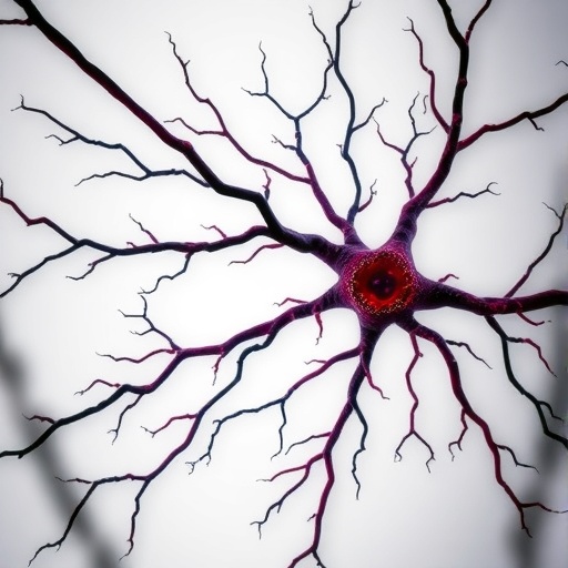
PV⁺ interneurons are fast-spiking inhibitory cells that play critical roles in orchestrating neuronal network oscillations and synaptic integration in the hippocampus. Their function is essential for proper cognitive processes such as learning and memory. Previous studies have established that chemogenetic activation of a small population of these cells induces expression changes in various genes including Vgf, culminating in increased PV–PV connectivity. However, whether such regulation arises naturally during memory processes had remained elusive until now.
In the latest experiments, contextual fear conditioning—a widely used paradigm to induce associative fear memory through brief foot shocks—was employed to probe the physiological engagement of PV⁺ interneurons in the CA1 region of the hippocampus. Animals subjected to this single-trial conditioning reliably exhibited prolonged freezing behavior when re-exposed to the context, reflecting successful learning. At the cellular level, the activated subpopulation of PV⁺ interneurons was identified via FOS expression, a marker for recent neuronal activity, approximately two hours post-conditioning, confirming time-specific recruitment of these inhibitory cells.
Intriguingly, these cFC-activated PV⁺ interneurons were found to receive significantly fewer synapses from other PV⁺ cells compared to their inactive neighbors. This suggests that PV⁺ interneurons with lower baseline inhibitory input are preferentially mobilized during fear learning, pointing to a network-level mechanism where selective disinhibition liberates specific interneurons for engagement. These observations underscore the nuanced interplay between intrinsic connectivity and recruitment during memory encoding.
To circumvent the transient nature of FOS expression and better capture the identity of activated PV⁺ interneurons over extended periods, researchers employed a Cre-dependent robust activity marking (CRAM) system. This innovative genetic tool labels neurons based on activity-induced promoter activation, leading to sustained tdTomato expression that persists well beyond immediate early gene expression windows. Through CRAM, investigators distinguished PV⁺ cells engaged by cFC from their quiescent counterparts days after conditioning, enabling longitudinal analysis of plasticity markers and synaptic changes.
Subsequent analyses revealed a marked elevation of Vgf expression within these activity-tagged PV⁺ interneurons relative to non-activated neighbors at 24 hours post-conditioning. This sustained upregulation corroborates prior chemogenetic findings and highlights Vgf as a key mediator linking neuronal activation to molecular plasticity programs. Notably, Vgf—originally characterized as a neuropeptide precursor—is increasingly recognized for its roles in synaptic modulation and intracellular signaling pathways that govern inhibitory circuitry adaptation.
Researchers then explored whether the enhanced Vgf levels corresponded with alterations in PV–PV synaptic connectivity over time. Strikingly, although activated PV⁺ cells initially exhibited lower densities of inhibitory synapses received from fellow PV⁺ interneurons at 24 hours post-cFC (mirroring earlier observations at 2 hours), these synaptic contacts progressively increased over the subsequent 48 hours. This temporal pattern suggests a delayed but robust homeostatic strengthening of network inhibition targeting the active interneurons, likely orchestrated through Vgf-dependent mechanisms.
Such findings illuminate a feedback system wherein PV⁺ interneurons that escape strong initial inhibition become preferentially active during learning episodes and then undergo adaptive remodeling to recalibrate inhibitory drive. This plasticity may serve to fine-tune the balance of excitation and inhibition within hippocampal circuits, stabilizing network dynamics following behavioral activation and contributing to the consolidation of memory traces.
Furthermore, the data imply that neuropeptide signaling pathways, exemplified by Vgf induction, could be instrumental in governing interneuron-specific synapse formation and functional connectivity. The precise molecular cascades downstream of Vgf remain to be fully elucidated, but the evidence points to a pivotal role in interneuronal communication and synaptic refinement aligned with experiential demands.
These insights are not only pivotal for understanding hippocampal function but may have broader implications for disorders characterized by inhibitory circuit dysfunctions, such as epilepsy, schizophrenia, and autism spectrum disorder. Targeting molecules like Vgf or modulating PV–PV connectivity could emerge as novel therapeutic strategies aimed at restoring inhibitory balance in pathological states.
The integration of genetic labeling strategies, high-resolution synaptic imaging, and behavioral paradigms in this investigation exemplifies the synergistic approach necessary to decode the complexities of neural plasticity. By illuminating how neuropeptide-encoding genes regulate interneuron plasticity in vivo, this research advances our grasp of the cellular substrates underlying learning and memory.
As hippocampal interneurons are pivotal orchestrators of network oscillations and synchrony, understanding the modulation of their inhibitory synapses provides foundational knowledge that bridges molecular neurobiology with systems neuroscience. The adaptive increase in PV–PV connectivity following fear conditioning portrays a dynamic circuitry capable of self-regulation and structural remodeling in response to environmental stimuli.
In conclusion, this study elegantly demonstrates that contextual fear conditioning selectively recruits a subset of PV⁺ interneurons distinguished by low baseline inhibitory input, provoking increased expression of Vgf that ultimately enhances PV–PV synaptic connectivity. Such plastic changes unfold over days and represent an intrinsic mechanism by which inhibitory networks adjust during memory processes, enriching our conceptual framework of neuronal adaptability.
Subject of Research: Regulation of plasticity in parvalbumin-expressing (PV⁺) interneurons in the hippocampus during fear learning.
Article Title: Regulation of PV interneuron plasticity by neuropeptide-encoding genes.
Article References:
Selten, M., Bernard, C., Mukherjee, D. et al. Regulation of PV interneuron plasticity by neuropeptide-encoding genes. Nature (2025). https://doi.org/10.1038/s41586-025-08933-z
Image Credits: AI Generated
Tags: behavioral stimuli and neural adaptationcognitive processes in learning and memorycontextual fear conditioning effectsfast-spiking inhibitory neuronsfear learning mechanismshippocampal interneurons and memoryneuronal network oscillationsneuropeptide gene regulationparvalbumin-expressing interneuronsPV interneuron plasticitysynaptic connectivity in PV interneuronsVgf gene expression in neurons


