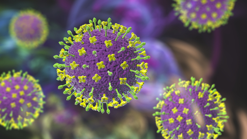
Nipah virus is a zoonotic virus harbored by fruit bats. It can be transmitted to pigs and humans, infect people through contaminated food, and can travel directly from person to person via droplets. In severe cases, the infection can cause serious respiratory illness and encephalitis. The virus kills between 40 and 75 percent of those infected, according to estimates from the Centers for Disease Control and Prevention. Currently, there are no vaccines to prevent or mitigate infection with the Nipah virus, and no effective treatments for the disease other than supportive care.
Now, researchers used cryoelectron microscopy (cryo-EM) to elucidate the structure of the Nipah virus polymerase complex (the large protein and phosphoprotein). Not only that, but they also performed structural, biophysical, and in-depth functional analyses of the polymerase.
This work is published in Cell in the paper, “Structural and functional analysis of the Nipah virus polymerase complex.”
The World Health Organization has declared Nipah virus a priority pathogen, a designation given to organisms that can cause serious outbreaks and require urgent research to inform prevention and treatment strategies.
Until now, the structure and function of the Nipah virus polymerase remained poorly understood. Unraveling this piece of the viral apparatus is the critical first step toward developing ways to combat a virus that poses a serious threat.
“Identifying how the polymerase is regulated to switch on and switch off the different enzymatic activities that are required for viral replication would be game-changing, and this study represents a key step towards that goal,” said Rachel Fearns, PhD, professor of virology, immunology & microbiology at the Boston University Chobanian & Avedisian School of Medicine.
Once the researchers worked out the structure of the enzyme, they took a closer look at how different parts of the enzyme affect the different functions that it performs. They found that “the L protein assembles with a long P tetrameric coiled-coil that is capped by a bundle of ⍺-helices that we show are likely dynamic in solution.” In addition, docking studies with a known L inhibitor clarify mechanisms of antiviral drug resistance.
They also identified “L protein features that are required for both transcription and RNA replication and mutations that have a greater impact on RNA replication than on transcription.”
“Elucidating both the unique and shared characteristics of the Nipah virus polymerases in comparison to other viral polymerases, our study provides critical insights that have the potential to inform the development of broad-spectrum antivirals,” said Heesu Kim, a technician in the Fearns Lab.
Nipah virus has the potential to ignite a pandemic, the researchers said, because it can spread via airborne droplets and respiratory secretions. The researchers also noted that there is one promising oral drug candidate developed by scientists at Georgia State University that works against viruses related to Nipah but not against Nipah virus itself. To understand why this drug candidate is ineffective against Nipah virus, the researchers conducted various simulation studies to see whether certain structural tweaks to the viral polymerase would improve the ability of the drug to latch onto the virus. The researchers identified a specific portion of the viral polymerase that could become a drug target. This in turn can inform the design of small-molecule inhibitors that disrupt the viral polymerase and render Nipah virus susceptible to treatment.
“This new understanding can help us identify the functional properties of the polymerase structure that could be leveraged as drug targets,” said Jonathan Abraham, PhD, associate professor of microbiology at Harvard Medical School and HHMI investigator.