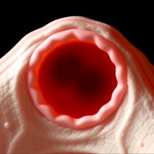
After ovulation, an egg begins a carefully timed journey through the oviduct, also known as the Fallopian tube, toward the uterus. Fertilization normally takes place within this passage, where the egg may encounter sperm and, if fertilization occurs, develop into an early embryo while continuing toward the womb. For decades, scientists have assumed that the distinctive longitudinal folds lining the oviduct play a central mechanical role in guiding this movement. New research from Japan now challenges that assumption, showing that eggs can still reach the uterus even when these folds lose their normal alignment.
The oviduct is lined by an epithelium containing multiciliated cells. Each of these specialized cells carries roughly 200 motile cilia, microscopic hair-like structures that beat in a coordinated manner. Their collective movement produces fluid flow and mechanical forces directed from the ovary toward the uterus. This directional transport is essential because mammalian eggs and early embryos cannot move independently through the oviduct. The epithelial surface is also shaped into prominent folds that normally extend along the same ovary-to-uterus axis, creating an intricate three-dimensional landscape inside the tube.
Longitudinal epithelial folds are not unique to mammals. Similar structures occur in the oviducts of birds, amphibians, and other vertebrates, suggesting that they may have been preserved through evolution because they provide an important reproductive advantage. Their parallel arrangement has led researchers to propose that the folds act like tracks, channels, or a conveyor system that helps direct eggs toward the uterus. Yet testing this idea has been difficult. Genetic changes that alter fold formation frequently affect other features at the same time, including the orientation of cilia, tissue shape, or the overall development of the oviduct.
A team led by Dr. Masaki Arata and Professor Toshihiko Fujimori at Japan’s National Institute for Basic Biology found a way to separate these factors. The researchers examined female mice with greatly reduced levels of VANGL1, a protein involved in planar cell polarity. Planar cell polarity coordinates the orientation of cells and structures across the surface of a tissue, rather than along its thickness. In normal mouse oviducts, epithelial folds run relatively straight from the ovarian end toward the uterus. In the VANGL1-deficient animals, however, the folds became irregular, misaligned, and extensively branched.
The structural disruption was striking, but it did not produce the expected failure of transport. Using high-speed microscopy and quantitative image analysis, the researchers found that the cilia in mutant oviducts continued to beat predominantly along the ovary-to-uterus axis. The animals were also fertile and produced offspring, demonstrating that eggs or early embryos could pass through the altered oviduct and arrive in the uterus. This result indicates that directional ciliary activity, rather than the geometric alignment of the epithelial folds alone, can provide the primary driving force for transport.
The researchers then tested movement more directly by placing fluorescent microspheres on the exposed inner surface of the oviduct. These beads served as visible tracers of the fluid and surface forces generated by the cilia. In both normal and mutant tissues, the beads generally moved toward the uterus. Their paths were not identical, however. In the mutant oviducts, movement was slower, trajectories were less linear, and beads were occasionally trapped near regions where the abnormal folds branched. The findings suggest that the folds are not essential for establishing direction, but they may reduce resistance, stabilize flow, or help prevent particles from becoming temporarily immobilized.
The distinction is important because fertility alone would have hidden these differences. The mutant mice were capable of reproduction, yet their internal transport system operated less efficiently. By combining fertility measurements with live imaging and quantitative tracking, the researchers showed that successful passage and optimal passage are not the same biological outcome. A tissue architecture can be dispensable in the strict sense—its loss does not prevent reproduction—while still improving the speed, consistency, and reliability of a vital physiological process.
“Our findings suggest that the longitudinal folds are not an indispensable conveyor belt for oocyte transport,” Professor Fujimori said. Instead, the folds may create a favorable structural environment in which ciliary forces can act more effectively. Dr. Arata noted that the mutant model was particularly informative because cilia retained much of their normal orientation while fold architecture was severely disrupted. This separation allowed the researchers to distinguish the contribution of epithelial geometry from that of the directional force generated by multiciliated cells.
The study, published in the Proceedings of the National Academy of Sciences, provides a more nuanced view of how the oviduct functions. Rather than relying on a single anatomical mechanism, the tissue appears to combine active transport generated by cilia with a structural design that improves performance. The results may also inform research on other ciliated organs, including the respiratory tract and brain ventricles, where coordinated ciliary motion operates within complex epithelial landscapes. Understanding how tissue architecture modifies ciliary transport could ultimately clarify why structural abnormalities sometimes impair physiological efficiency without causing complete organ failure.
Subject of Research: The role of longitudinal epithelial folds and multiciliated cells in transporting oocytes through the mouse oviduct.
Article Title: Successful oocyte transport through the oviduct does not depend on the longitudinal alignment of oviduct epithelial folds
News Publication Date: 4 August 2026
Web References: https://doi.org/10.1073/pnas.2605383123
References: Proceedings of the National Academy of Sciences; DOI: 10.1073/pnas.2605383123
Image Credits: Division of Embryology, National Institute for Basic Biology (NIBB)
Keywords: oviduct, Fallopian tube, oocyte transport, cilia, multiciliated cells, epithelial folds, planar cell polarity, VANGL1, reproductive biology, mouse development, tissue architecture, embryo transport
Tags: embryo transport in fallopian tubesevolutionary conservation of oviduct structuresfallopian tube cilia functionimplications for fertility and reproductive healthinfluence of epithelial folds on fertilizationmammalian egg movementmotile cilia in oviductoocyte transport mechanismsoviduct epithelial foldsreproductive tract structural featuresrole of ciliated epithelial cellstubal epithelial morphology