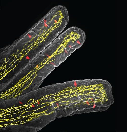
![A 3D view inside the small intestine showing its finger-like projections called villi. Red cells release serotonin, a chemical messenger that triggers sensations like nausea. Yellow fibers are nerve endings that detect these signals and send them to the brain. [Koki Tohara/UCSF]](https://www.genengnews.com/wp-content/uploads/2026/03/Low-Res_Nature-villi-image-e1774471673972.jpg "Low-Res_Nature villi image")
Anyone who has weathered a bad stomach bug knows the feeling: a loss of appetite that sets in and lingers, even after the initial illness. For the millions of people around the world who are chronically infected with parasitic worms, the same thing happens. But scientists have long puzzled over exactly why.
Researchers at UC San Francisco now report that they have traced the molecular pathway that connects the gut immune system to the brain during a parasitic infection, explaining how the immune system triggers a loss of appetite.
The findings reveal an unexpected communication system between two cell types, tuft cells and serotonergic enterochromaffin cells (ECs), and could shed light on a range of conditions involving gut discomfort—from food intolerances to irritable bowel syndrome.
“The question we wanted to answer was not just how the immune system fights parasites, but how it recruits the nervous system to change behavior,” said co-senior author David Julius, PhD, professor and chair of Physiology at UCSF and recipient of the 2021 Nobel Prize in Physiology or Medicine. “It turns out there’s a very elegant molecular logic to how that happens.” Julius is senior author of the researchers’ published paper in Nature, titled “Parasites trigger epithelial cell crosstalk to drive gut–brain signaling,” in which they concluded, “Our investigation of paracrine communication between tuft and EC cells now reveals a direct link between sensory and immune systems that alters food intake through the gut–brain axis.” The UCSF researchers worked in collaboration with Stuart Brierly, PhD, and his lab group at the University of Adelaide in Australia.
“Parasitic infections modulate both immune and sensory responses, but how these systems collaborate to elicit protective behaviors remains incompletely understood,” the authors wrote. “Gastrointestinal symptoms during parasite infection have been vaguely attributed to tissue damage and alterations in the gut microbiota.”
The epithelial lining of the gut contains specialized sensory cells, including enterochromaffin cells and tuft cells, that detect and act as first lines of defense against pathogens and irritants, the team further explained. Tuft cells detect parasites and trigger immune defenses, while enterochromaffin cells release signals that activate nerve fibers leading to the brain. EC cells are known to cause sensations like nausea, pain, and gut discomfort, but whether they communicate with tuft cells was unknown. “If tuft and EC cells collaborate to detect noxious stimuli, they should be capable of communicating through some sort of paracrine signaling mechanism,” they stated.
“My lab has long been interested in how tuft cells, after they initially respond to a parasitic infection, release signals to other cell types,” said co-senior author Richard Locksley, MD, a UCSF immunologist.
First author Koki Tohara, PhD, a postdoctoral researcher at UCSF, found the answer by positioning genetically engineered sensor cells directly next to tuft cells under a microscope. When tuft cells were exposed to succinate, a molecule produced by parasitic worms, the sensor cells lit up, revealing that tuft cells were releasing acetylcholine (Ach), a chemical messenger used primarily by neurons.
When acetylcholine was added to lab-grown gut tissue containing EC cells, they released serotonin. This activated vagal nerve fibers that carry signals from the gut to the brain. “What we found is that tuft cells are doing something neurons do, but by a completely different mechanism,” Tohara said. “They’re using acetylcholine to communicate, but without any of the usual cellular machinery that neurons rely on to release it.”
The team also discovered that tuft cells release acetylcholine in two distinct phases, explaining why people often don’t develop a loss of appetite until days into an infection. In the first phase, a brief burst of acetylcholine is released. Later, after the immune system has mounted a full response, tuft cells multiply and produce a slow, sustained release of acetylcholine that is sufficient to activate EC cells. “We find that tuft cells use two distinct mechanisms of acetylcholine(ACh) release …” they wrote.
“These include acute release in response to parasite-derived metabolites, followed by constitutive ‘leak-like’ release, which occurs with type 2 inflammation.” And it’s only the sustained mode of ACh release that elicits levels of serotonin sufficient to stimulate vagal afferent neurons that suppress food intake. “This two-phase paracrine signaling mechanism explains how parasitic infection progresses from an initial asymptomatic phase to symptomatic established disease, in which type 2 immune and sensory signaling pathways within the gut–brain axis collaborate to evoke protective behaviors.”
Julius noted, “This explains why you feel fine at first but then start to feel sick as the infection becomes established. “The gut is essentially waiting to confirm that the threat is real and persistent before it tells the brain to change your behavior.”
To test whether the pathway matters beyond the lab, the researchers infected mice with a parasitic worm and tracked their food intake. Mice with normal tuft cell function ate less as the infection took hold. Mice engineered to lack acetylcholine-producing machinery in their tuft cells kept eating normally, confirming that the molecular chain drives the behavioral response. The new findings could have relevance for treating the symptoms of a parasite infection.
“Controlling the outputs of tuft cells could be a way to control some of the physiologic responses associated with these infections,” Locksley said, adding that the study also could have broader implications.
Tuft cells are found throughout the body—not just in the gut, but also the airways, gallbladder, and reproductive tract—and disruptions to the newly identified pathway could contribute to conditions like irritable bowel syndrome, food intolerances, and chronic visceral pain.