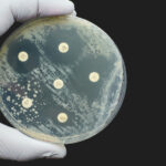

Errors in chromosome segregation cause more than 80% of early human embryos to contain cells with an incorrect number of chromosomes, a phenomenon called aneuploidy. While aneuploid cells are normally depleted from embryonic germ layers to give rise to healthy births, the emergence of aneuploidy in somatic tissues is associated with diseases, such as cancer, with 90% of human solid tumors reported to be aneuploid. Understanding how aneuploid cells are eliminated during these early developmental stages is crucial for gaining insights into fertility and developing cancer treatments.
In a new study published in Cell Genomics titled “Depletion of aneuploid cells is shaped by cell-to-cell interactions,” researchers from the Institute for Research in Biomedicine (IRB) Barcelona have developed a tool that can generate and label customized aneuploid cells in living tissue. The new tool offers a window through which to observe the behavior of these cells in real-time.
To date, the most popular technique to induce aneuploidy and analyze its impact on disease has been by triggering chromosome mis-segregation events in dividing cells. However, this approach does not allow selection of the chromosome sets that are being generated.
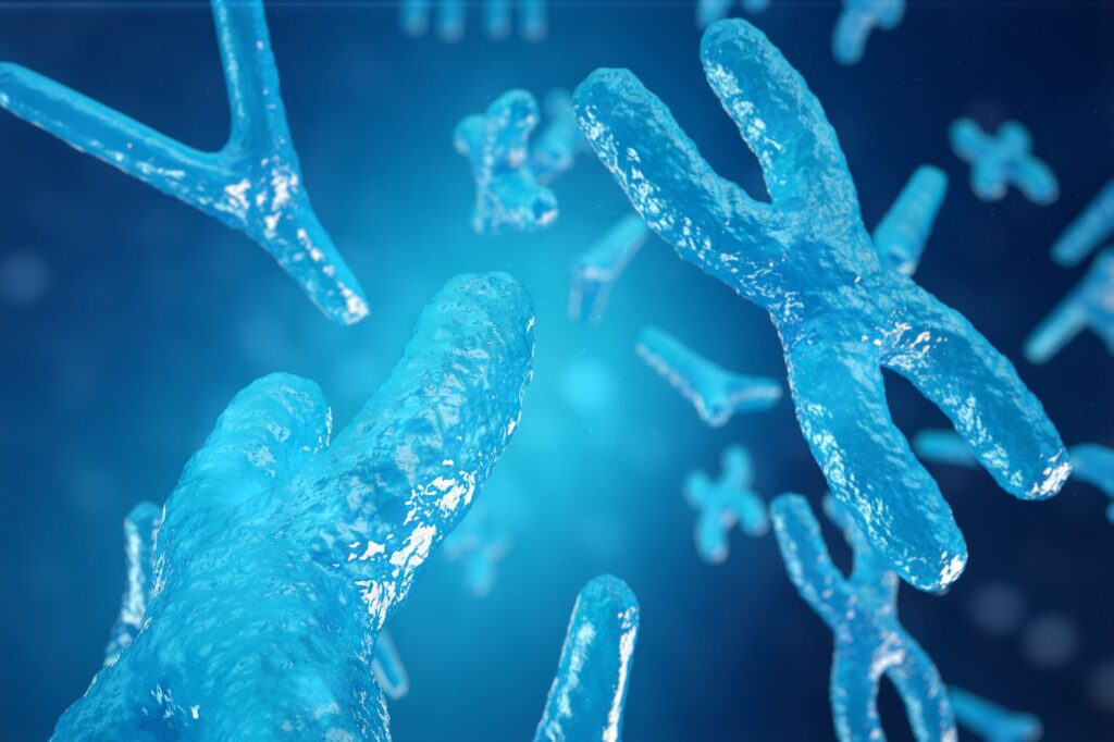
Using Drosophila as a model system, the authors applied a tool based on the FLP-FRT recombination system, a site-specific recombination technology used to manipulate DNA in vivo, to generate both monosomies, a single chromosome copy, and trisomies, three chromosome copies. As monosomic cells lose one of the two chromosomal doses, some genes become haploinsufficient, where a single copy no longer produces enough protein to keep the cellular machinery operating at full capacity.
Among the most well-known haploinsufficient genes are those that code for ribosomal proteins, the fundamental building blocks of the cell’s protein-making machinery. Reduction of a single ribosomal subunit can lead to a significant slowdown of protein production, leading to increased cellular stress.
The authors generated monosomic cells within normal tissue and demonstrated that the genome contains a large number of haploinsufficient genes beyond those coding for ribosomal proteins. Results showed that monosomic cells are eliminated through various molecular mechanisms of cell competition.
The study also generated monosomic and trisomic cells simultaneously in the same tissue and observed that the latter can accelerate the removal of monosomic cells. According to Elena Fusari, PhD, a researcher at IRB Barcelona and first author of the study, the “fittest” cells push aneuploidies toward apoptosis, while aneuploid cells that were left alone were shown to survive.
“In the field of assisted reproduction, there’s a growing reconsideration of current embryo selection criteria. This shift comes as new research suggests that embryos may actually be capable of eliminating problematic cells on their own,” said Fusari.
The results highlighted the importance of cell-to-cell interactions, which paves broader implications for treatments that modify neighboring cells to force the removal of pathological clones. Understanding the “rules” of competition between aneuploid cells also presents a path to develop therapies for cancer cells.
As next steps, the researchers plan to carry out an exhaustive search of all haploinsufficient regions of the Drosophila genome with the goal of mapping which genes trigger competition signals. Modulating this response could increase the success rate of assisted reproduction treatments and develop drugs to tackle aneuploidy, which is characteristic of many tumors.