
Scientists have traditionally thought of the brain as a single, unified organ. But new research by Stanford Medicine scientists, working with collaborators California Institute of Technology and the University of California, San Francisco, indicates that what we call the brain is two distinct organs that evolved independently over hundreds of millions of years.
The discovery overturns a prevailing model of brain development, based on the theory that there is a single progenitor cell early in development that gives rise to the entire brain. This model suggested that all parts of the brain shared a common developmental origin. The newly reported findings show that the human brain consists of two ancient nervous systems cleverly packaged together—a more primitive part that regulates the heart beating, our breathing and other functions, and another part that makes us distinctly human, capable of poetry, mathematics and wondering about our own origins.
The results could help explain why scientists have struggled for decades to grow certain types of brain cells in the laboratory—and it opens new avenues for studying devastating diseases that affect the brain stem, such as spinal muscular atrophy (SMA) and amyotrophic lateral sclerosis (ALS).
“We’ve shown for the first time that the front of the brain arises from a totally different progenitor cell than the back of the brain,” said Kyle Loh, PhD, associate professor of developmental biology. “Our discovery means that we can now grow neurons from the back of the brain, the hindbrain, in a petri dish and study their functions.” Loh is senior author, and Carolyn Dundes, PhD, and Rayyan Jokhai are co-first authors of the scientists’ published paper in Nature Neuroscience, titled “Two parallel neural ectoderm progenitors contribute to the developing brain,” in which they concluded that “… the emerging notion of two parallel brain progenitors has a number of ramifications for development, differentiation and evolution.”
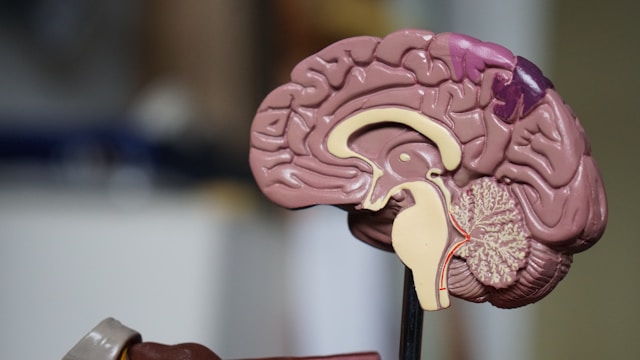
The adult brain has three main regions: the forebrain, midbrain and hindbrain. The forebrain handles higher-level thinking—language, consciousness and abstract reasoning. In contrast, the hindbrain, located at the back of the skull and often called the brain stem, controls essential, automatic functions that keep us alive: breathing, sleeping, and regulating our heartbeat and hunger urges. “Consequently, hindbrain injury and diffuse intrinsic pontine glioma (a childhood hindbrain cancer) are both deadly, as they impair consciousness, sensation, reflexes and breathing,” the authors wrote. The hindbrain neurons also control the muscles of the face, tongue and throat, which affect speech and swallowing.
Despite the critical importance of the hindbrain, scientists have struggled for decades to generate human hindbrain neurons in the laboratory. This gap has hampered research into devastating diseases affecting the brain stem, including spinal muscular atrophy and amyotrophic lateral sclerosis.
SMA is a leading genetic cause of death in children under one year of age. ALS, which is often diagnosed between the ages of 40 years and 70 years of age, affects both the forebrain and the hindbrain. In both disorders, certain hindbrain neurons gradually cease to function, and the patient loses the ability to swallow, which can cause pneumonia when food or liquid is inhaled into the lungs; eventually, patients lose the ability to breathe. “… degeneration of hindbrain motor neurons probably compromises eating and swallowing in diseases such as spinal muscular atrophy and amyotrophic lateral sclerosis, leading to choking, aspiration, pneumonia and, in some cases, death,” the team continued.
The researchers’ newly reported discoveries emerged from studying the earliest moments of embryonic development, during a stage called gastrulation when the body first takes shape. Jokhai and Dundes discovered that the hindbrain follows a separate developmental path, running in parallel to, rather than branching off from, the pathway that creates the forebrain and midbrain.
The researchers learned this from examining developing mouse embryos. They identified two different brain progenitor cells. “Two parallel brain progenitors emerge simultaneously during gastrulation: anterior neural ectoderm (forebrain/midbrain progenitor) and posterior neural ectoderm (hindbrain progenitor),” they stated. One, which expresses a gene called Otx2, is destined to become the forebrain and midbrain. The other, which expresses a gene called Gbx2, is committed to forming the hindbrain. They showed that these two cell populations never overlap; they are mutually exclusive from the earliest stages of development. “Differentiation of human pluripotent stem cells into anterior or posterior neural ectoderm-like cells revealed these were lineage committed to forebrain/midbrain versus hindbrain fates, respectively.”
The team then examined the DNA packaging, or chromatin, in these cells. Chromatin is a way cells determine which genes can be easily accessed and which are bundled away out of reach. What they found was striking: The anterior neural ectoderm (aNE; future forebrain and midbrain) and posterior neural ectoderm (pNE; future hindbrain) have fundamentally different chromatin configurations. “They harbored diverging chromatin landscapes foreshadowing future forebrain/midbrain versus hindbrain identities,” the team noted. These differences essentially locked each progenitor cell into its respective fate, like travelers on parallel tracks that never cross.
This revelation explained decades of frustration in the field—scientists had been trying to turn one type of progenitor cell into another that it is fundamentally incapable of becoming. “Previous attempts to make hindbrain neurons likely tried to coax forebrain and midbrain progenitors into hindbrain cells, which our study shows is not possible,” Jokhai said. “In stem cell biology, people are always fixated with creating the end cell type, like the neuron. But it’s important to begin at the earliest stages of embryonic development. Our careful attention to that early time point allowed us to find this fundamental split in brain development.”
Armed with this knowledge, the researchers for the first time successfully coaxed human pluripotent stem cells (a kind of cell that can create any cell in the human body) to become functional hindbrain motor neurons in the laboratory.
These lab-grown neurons displayed all the hallmarks of authentic hindbrain cells: They exhibited waves of electrical activity called action potentials and made proteins that identify the segments of the hindbrain that control facial and swallowing muscles. “Discovering this lineage bifurcation provided a roadmap to differentiate hPS cells into pNE and subsequently specific types of hindbrain neuron in vitro, thus enabling future explorations of human hindbrain biology and disease,” the scientists noted.
Finally, the researchers looked back over 550 million years of evolutionary time. They found the same two-origin brain pattern in chickens, zebrafish and, remarkably, in acorn worms, tiny creatures living on the ocean floor that share a distant common ancestor with humans. “Separate anterior and posterior ectoderm populations arise during gastrulation across deuterostome species as diverse as acorn worm, zebrafish, chicken, mouse and primate, implying that this distinction between two different types of ectoderm predates the origins of chordates and arose ~550–600 million years ago,” the team further stated. Jellyfish, which diverged from humans about 600 to 700 million years ago, have two nervous systems at different ends of their body.
“Our research suggests that evolution took two existing neural systems and pushed them together spatially,” Loh said. “Having the brain as one organ would probably be more efficient, but we rely on this primordial way to make the brain as two separate pieces.”
“I was surprised at our findings because the word ‘brain’ implies a contiguous organ that likely has a singular origin,” Jokhai added. “But even 500 million years ago, there were these separate neural systems, which now almost operate as one, which is very cool.”
The research also has implications for investigating treatments for SMA, ALS and other conditions affecting the brain stem. Until now, studying these diseases has been nearly impossible because scientists cannot obtain brain stem tissue from living patients. The ability to grow these neurons in a dish opens new possibilities for understanding what goes wrong. There’s even an unexpected connection to obesity treatment: The hindbrain contains circuits that regulate hunger—which is precisely how weight-loss drugs like semaglutide work.
The researchers would like to extend their studies to determine the developmental origins of the spinal cord and to learn exactly how SMA and ALS compromise the function of hindbrain neurons. “Now we have a model to better understand these devastating diseases, and work toward regenerative therapies for them,” Jokhai said. “This is a very exciting new frontier in brain research.”