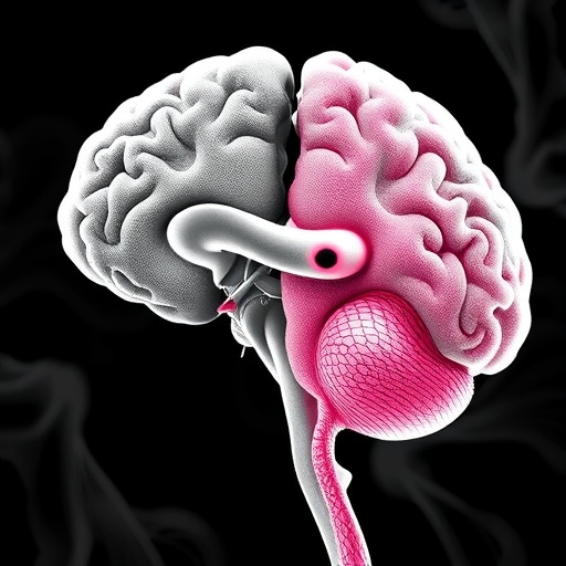
In recent years, the scientific community has devoted considerable effort to unraveling the complex mechanisms underpinning neurological disorders, particularly those involving white matter abnormalities detectable via magnetic resonance imaging (MRI). A groundbreaking study published in Nature Communications in 2026 by Parent, Alasmar, Osborne, and colleagues offers a detailed and nuanced exploration of white matter hyperintensities (WMHs), a radiological hallmark frequently associated with aging, vascular disease, and neurodegeneration. This research provides unprecedented insight into the spatiotemporal pathophysiology of WMHs directly within the living brain, marking a significant advance in our understanding of their vascular and neurodegenerative origins.
White matter hyperintensities, visible as bright spots on T2-weighted MRI scans, have been long acknowledged as predictors of cognitive decline, stroke risk, and dementia. However, the precise biological processes giving rise to these lesions remain debated. Traditionally, WMHs have been viewed primarily through the lens of small vessel ischemia, where chronic hypoperfusion leads to demyelination and axonal damage. Alternatively, emerging hypotheses propose a neurodegenerative component, wherein proteinopathies and neuronal loss also contribute to white matter damage. The study by Parent et al. innovatively integrates advanced neuroimaging techniques with computational modeling to disentangle these overlapping pathological processes in vivo.
Employing longitudinal MRI datasets spanning several years, the authors meticulously charted the progression of WMHs across multiple brain regions while simultaneously assessing cerebral blood flow and markers of neurodegeneration. Their use of sophisticated spatiotemporal mapping allowed them to discern patterns of lesion development previously obscured in cross-sectional analyses. Notably, they identified that early-stage WMHs often localize to watershed areas prone to hypoperfusion, consistent with vascular etiology. However, as lesions enlarged and expanded into deep white matter, neurodegenerative signatures such as cortical thinning and tau pathology emerged as dominant contributors.
This dual-pathway model proposed by the researchers challenges the traditional dichotomy that strictly classifies WMHs as either vascular or neurodegenerative. Instead, it reveals a dynamic interplay wherein initial vascular insults set the stage for subsequent neurodegenerative processes, creating a vicious cycle accelerating white matter damage. Such revelations have significant implications for clinical practice and therapeutic strategies. Targeting vascular risk factors alone may not suffice, especially in later stages where neurodegeneration becomes prominent. A combination of vascular health optimization and neuroprotective interventions may be required to halt or slow disease progression effectively.
The methodology adopted by the team incorporated advanced MRI sequences sensitive to microstructural integrity, such as diffusion tensor imaging (DTI) and arterial spin labeling (ASL), providing both structural and perfusion data. These imaging modalities enabled precise quantification of tissue damage and blood flow deficits with unparalleled resolution. Furthermore, the integration of machine learning algorithms facilitated the classification of WMHs into subtypes based on their growth trajectories and spatial distribution, paving the way for personalized medicine approaches in neurology.
One of the study’s remarkable findings relates to the temporal dynamics of white matter lesions. By analyzing changes over multiple follow-up points, Parent et al. detailed how early microvascular dysfunction leads to subtle white matter changes, which in turn predispose these regions to accumulate pathological proteins characteristic of neurodegeneration. The timing and sequence of these events bear critical importance in identifying therapeutic windows for intervention and prognosticating patient outcomes in conditions like vascular dementia and Alzheimer’s disease.
The authors also shed light on the regional variability of WMH pathology. While periventricular WMHs showed a stronger link to vascular factors, deep white matter lesions correlated more closely with neurodegenerative markers. This spatial heterogeneity underscores the importance of region-specific assessments rather than relying solely on global WMH burden. Such granular analysis enhances diagnostic specificity and informs risk stratification for patients with mild cognitive impairment or at prodromal stages of dementia.
Implications extend beyond clinical diagnosis and treatment. From a research perspective, the study advocates for a paradigm shift in how neurologists and neuroscientists conceptualize white matter lesions. It compels the field to move towards integrative frameworks that consider multifactorial origins and evolving pathologies rather than simplistic cause-effect models. This new understanding also encourages cross-disciplinary collaborations spanning vascular biology, neuroimaging technology, computational modeling, and neurodegenerative disease research.
Additionally, the study underscores the power of in vivo human imaging combined with computational analysis as a transformative tool in neurobiology. Traditionally, much knowledge about white matter pathology derived from post-mortem studies and animal models, which have inherent limitations in capturing dynamic disease processes. Real-time longitudinal imaging of living human brains, as demonstrated here, offers unprecedented opportunities to observe disease evolution and test hypotheses directly within the clinical context.
The broader societal impact of these findings cannot be overstated. As the global population ages, the burden of dementia and cerebrovascular diseases is set to rise dramatically. White matter hyperintensities are highly prevalent in elderly individuals, making them a critical target for early diagnosis and intervention. By elucidating the complex pathophysiology of WMHs, this research lays the groundwork for developing biomarker-driven trials, novel therapeutics, and personalized healthcare strategies aimed at preserving cognitive health in aging populations.
Moreover, the study’s insights may inform public health initiatives emphasizing vascular health as a modifiable factor in neurodegeneration prevention. Lifestyle interventions promoting cardiovascular fitness, blood pressure control, and metabolic health gain renewed importance in light of their potential to influence white matter integrity and delay cognitive decline.
Importantly, while the study represents a major leap forward, the authors acknowledge remaining challenges and future directions. Validation in larger, diverse cohorts will be necessary to generalize findings and refine subtype classifications. Integration of additional biomarkers, such as cerebrospinal fluid profiles and genetic risk factors, could further enhance the mechanistic understanding of WMH pathophysiology. Moreover, translating these insights into effective clinical interventions remains a long-term goal necessitating interdisciplinary collaboration.
In sum, the work of Parent and colleagues marks a milestone achievement in neuroimaging and neurological research. By characterizing the spatiotemporal dynamics of white matter hyperintensities in vivo, and importantly, disentangling the vascular and neurodegenerative contributions, they provide an integrated framework that reshapes existing paradigms, informs clinical practice, and inspires future scientific endeavors. This study exemplifies how cutting-edge imaging technology combined with analytical rigor can illuminate the enigmatic processes underlying brain aging and neurodegenerative diseases, holding promise for better diagnosis, treatment, and ultimately prevention.
Subject of Research: Characterization of white matter hyperintensity pathophysiology in vivo, with a focus on distinguishing vascular and neurodegenerative contributions.
Article Title: Characterizing spatiotemporal white matter hyperintensity pathophysiology in vivo to disentangle vascular and neurodegenerative contributions.
Article References:
Parent, O., Alasmar, Z., Osborne, S. et al. Characterizing spatiotemporal white matter hyperintensity pathophysiology in vivo to disentangle vascular and neurodegenerative contributions. Nat Commun (2026). https://doi.org/10.1038/s41467-026-70832-2
Image Credits: AI Generated
Tags: advanced neuroimaging techniques in neurologycognitive decline biomarkers MRIcomputational modeling in brain researchdemyelination and axonal damage mechanismslongitudinal MRI studies of aging brainneurodegenerative causes of white matter changesproteinopathies in neurodegenerationsmall vessel ischemia and white matterspatiotemporal pathophysiology of WMHsstroke risk and white matter abnormalitiesvascular contributions to white matter damagewhite matter hyperintensities MRI