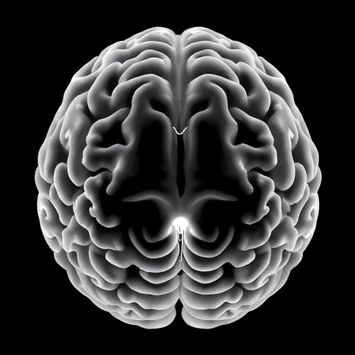
Researchers studying brain aging have uncovered evidence that cognition in older adults may depend on more than the preservation of gray matter alone. A study led by scientists at the Mark and Mary Stevens Neuroimaging and Informatics Institute at the Keck School of Medicine of USC suggests that superficial white matter—the thin layer of short-range nerve fibers directly beneath the brain’s outer surface—works together with gray matter to support mental abilities. The findings indicate that the condition of these local neural connections may influence how strongly gray matter loss affects cognition, potentially helping explain why people with similar levels of brain atrophy can experience very different cognitive outcomes.
Published in Alzheimer’s & Dementia: The Journal of the Alzheimer’s Association, the research analyzed brain scans and cognitive assessments from 459 adults aged 60 and older living in communities across India. The participants were part of the Harmonized Diagnostic Assessment of Dementia for the Longitudinal Aging Study in India, known as LASI-DAD. The study is notable not only for examining superficial white matter in detail, but also for drawing data from a population often underrepresented in neuroimaging research. More than half of the broader LASI-DAD sample has low literacy, approximately 60 percent live in rural communities, and many participants had little or no formal education.
Gray matter has traditionally dominated research into brain aging and dementia. It contains many of the neuronal cell bodies responsible for processing information, and reductions in gray matter volume, commonly referred to as atrophy, are strongly associated with cognitive decline. Yet the brain’s information-processing capacity also depends on communication between regions. Superficial white matter consists of short, curved fibers that link nearby areas of the cerebral cortex. These fibers form a local communication network, allowing neighboring regions to exchange signals rapidly. In a simple analogy, gray matter functions as a collection of processing centers, while superficial white matter provides the short roads that connect them.
To investigate the microscopic health of these pathways, the researchers used an advanced diffusion magnetic resonance imaging technique. Diffusion MRI tracks the movement of water molecules through tissue, revealing features that are not visible in conventional structural scans. The team focused on measures associated with neurite density and free water. Neurites are the small projections, including axons and dendrites, through which nerve cells communicate. A lower apparent neurite density may indicate a reduction or disruption of these cellular structures, while higher levels of freely moving water can reflect tissue changes such as loss of myelin, inflammation, swelling, or other forms of microstructural damage. Together, these measurements provided a more detailed view of superficial white matter integrity.
The participants also completed assessments covering language, memory, executive function, and visuospatial abilities. Across these domains, healthier superficial white matter was most consistently associated with stronger language performance. The clearest relationships appeared in frontotemporal regions, areas involved in recognizing words, producing fluent speech, and temporarily maintaining language-related information. These results do not mean that superficial white matter acts independently of gray matter. Instead, they suggest that the efficiency of local connections may help determine how well nearby cortical regions can work together, particularly when the tissue responsible for processing information has begun to deteriorate.
Gray matter atrophy remained the strongest overall predictor of cognitive performance in the analysis. However, its relationship with cognition changed depending on the state of the surrounding superficial white matter. When the local wiring showed poorer integrity, gray matter loss was more closely linked to weaker language abilities and greater cognitive impairment. When superficial white matter appeared healthier, the association between gray matter atrophy and poor performance was weaker. This pattern suggests a possible buffering effect: preserved short-range connections may help the brain continue coordinating activity even when some gray matter has been lost.
The researchers describe this possibility as a potential source of cognitive resilience, but they caution that the study cannot establish that healthy superficial white matter directly prevents decline. The analysis captured participants at a single point in time, meaning it cannot show whether superficial white matter changes occur before gray matter loss, after it, or alongside it. It also cannot determine whether improving the condition of these pathways would preserve cognition. Longitudinal studies that repeatedly scan the same individuals will be needed to track how gray matter and superficial white matter change together and to determine whether one tissue predicts future changes in the other.
The findings were particularly pronounced among participants who could not read or read incorrectly, had no formal education, or lived in rural areas. The researchers stress that these social conditions should not be interpreted as direct causes of brain damage. Rather, they may reflect the cumulative influence of lifelong educational opportunities, health care access, vascular risk, environmental exposures, occupation, nutrition, and other experiences that can shape brain aging. The stronger association between superficial white matter and language in these groups highlights the importance of studying cognition in populations with diverse social and educational histories, rather than assuming that results from highly educated urban populations apply universally.
Future research will examine whether vascular health, inflammation, Alzheimer’s-related proteins, and other biological factors help explain differences in superficial white matter integrity. The study’s authors also emphasize that brain aging is unlikely to be determined by a single structure or biomarker. A more complete picture may require measuring gray matter, white matter, blood flow, immune activity, protein accumulation, and lived environmental conditions together. By identifying superficial white matter as a potentially important partner of gray matter, the research opens a new avenue for understanding why cognitive aging varies so widely between individuals—and why protecting the brain’s smallest connections could prove as important as preserving its larger processing regions.
Subject of Research: People
Article Title: Superficial white matter and gray matter jointly support cognition among older adults in India
Web References: https://doi.org/10.1002/alz.71697; https://ini.usc.edu/
References: Alzheimer’s & Dementia: The Journal of the Alzheimer’s Association, DOI: 10.1002/alz.71697
Image Credits: Stevens INI
Keywords: Brain aging, superficial white matter, gray matter, cognition, language, dementia, Alzheimer’s disease, neuroimaging, diffusion MRI, cognitive resilience, older adults, India
Tags: Alzheimer’s disease and brain tissue analysisbrain agingbrain atrophy and individual cognitive differencescognitive assessment in aging populationseffects of local neural connections on cognitiongray and white matter in cognitionneural connectivity and cognitive declineneuroimaging research on older adultssuperficial white matter and cognitive healthunderrepresented populations in neuroimagingUSC brain aging studywhite matter and gray matter interaction