
According to some estimates, stroke is the second leading cause of death globally. One of the causes of a severe type of stroke are brain aneurysms. Now data from a new study suggests that certain cells in the brain may cause aneurysms to weaken and rupture. And it helps explain why some aneurysms burst while others do not. It also opens a door to new ways of potentially predicting and preventing strokes. All of the findings are covered in a new Nature Neuroscience paper titled “Cerebrovascular vulnerability and fibrosis in human brain aneurysms.”
Brain aneurysms, which are bulges in blood vessels in the brain, can go unnoticed for years before rupturing causing a severe, often deadly type of stroke. About one in 50 people in the U.S. has a brain aneurysm but predicting which ones are most dangerous remains challenging. Aneurysms can be repaired surgically or using other minimally invasive procedures but those decisions depend on the size and location of the aneurysm as well as patient specific risk factors. With the current study, “we’ve made major steps toward solving the mystery of how aneurysms form,” said Ethan Winkler, MD, PhD, assistant professor of neurological surgeon and senior author of the Nature Neuroscience study. “We’ve identified the cast of characters involved and seen which ones are implicated at different phases of disease.”
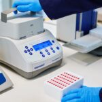
To get to those answers, Winkler and his team analyzed more than 100,000 individual cells from human aneurysms and healthy brain arteries. From these data, they identified 19 transcriptionally distinct cell types and determined which genes were active in each. They also mapped how the cells were organized within the blood vessel wall.
“Our atlas of human brain aneurysms, as well as cell-resolution spatial transcriptomics, revealed that pathological cerebrovascular remodeling occurs with the loss of structurally supportive smooth muscle cells and the emergence of activated perivascular fibroblasts, which re-populate the vascular wall and express multiple genes linked to aneurysm risk,” the scientists wrote.
Specifically, they found that vessels in aneurysm tissue had disorganized layers, and that many of the smooth muscle cells that allows the vessel walls to expand and contract had disappeared. In their place were scar-forming fibroblasts, which the team dubbed “activated fibroblasts.” These stiffened the arterial wall, making it less able to flex as blood flowed through. These cells also expressed genes that are linked to an inherited risk of aneurysm. The scientists also identified a type of macrophage that accumulated inside the arterial wall near the fibroblasts. The data showed that these specialized macrophages express a gene that is typically associated with bone tissue.
Further testing revealed the presence of a feedback look between the two cell types. Specifically, the activated fibroblasts release a signal that triggers the macrophages to produce enzymes that degrade the blood vessel’s structural support. The scientists confirmed that this was the case by blocking the signals sent to the macrophages. They observed that the macrophages were less likely to produce the destructive enzymes when the signal was blocked.
This process where vessel walls lose muscle cells followed by the buildup of scar tissue and immune cell activation helps explain why smaller aneurysms, which are often considered low risk, can still rupture. It jibes with Winkler’s own clinical experiences. He noted that more than half of the ruptures that he treated early in his career occurred in aneurysms below the typical surgical threshold of seven millimeters.
This study brings scientists and clinicians one step closer to understanding how aneurysms form and perhaps being able to intervene earlier to prevent them. As the scientists note in the paper, “the molecular blueprint provided by this study substantially extends our mechanistic understanding of brain aneurysms and nominates new cells and pathways with translational promise for the development of therapeutic options.” This could involve blocking the signals that fibroblasts send or by inhibiting the immune response to those signals.