
A new single-protein analysis technique gives researchers newfound ability to study scramblases, physiologically important proteins that translocate phospholipids bidirectionally across cell membranes.
In the study published in Nature Structural & Molecular Biology titled, “A single-vesicle fluorescence microscopy platform to quantify phospholipid scrambling,” researchers from Weill Cornell Medicine and Ruhr University Bochum have developed a fluorescence imaging-based technique to measure the activity rates of individual scramblase proteins.
“I’m excited about this new platform as it is versatile and provides unprecedented information on exactly how fast a single scramblase works,” said Anant Menon, PhD, professor of biochemistry and biophysics at Weill Cornell Medicine and co-corresponding author of the study.
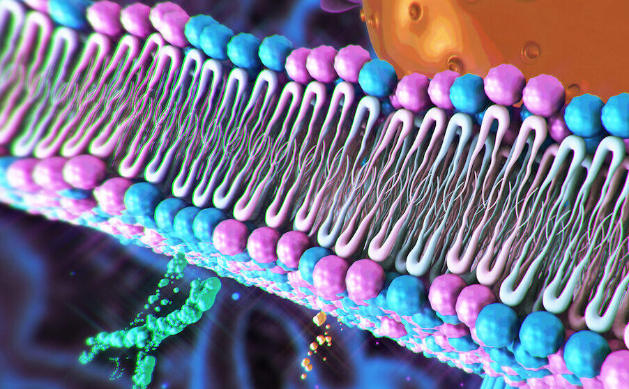
Scramblases are key drug targets with roles in the assembly of cell membranes, modification of proteins with sugars, cell survival, muscle development and molecular trafficking. Yet, strategies for understanding scramblase dynamics have been limited.
Traditionally, researchers purify scramblase proteins for further study using vesicles to record average scramblase activity. However, this bulk approach is unable to measure the transport rate of individual scramblases and capture how scramblase variability impact biological processes.
The authors used fluorescently-tagged scramblases to achieve high resolution and evaluated a scramblase protein, known as VDAC1, best known as a membrane channel protein within mitochondria. Two copies of VDAC must align to provide a pathway for lipid movement. These dimers have a wide range of scrambling rates, from fewer than 100 to more than 1,000 lipids per second.
“These findings indicate that only certain dimer conformations are capable of rapid scrambling, directly validating predictions from computer simulations,” Menon said.
The team demonstrated the versatility of their approach by applying the platform to measure lipid-scrambling by opsin, a cell-membrane receptor and scramblase that is involved in light-detection in the eye. Results showed that individual opsin proteins scramble lipids faster than VDAC dimers, achieving rates in excess of 10,000 lipids per second.
The new platform can study how drug molecules impact scramblase function. Additionally, the authors aim to combine their functional studies of scramblases with high-resolution imaging to understand how scramblase shape relates to activity rates. The team also plans to use the technique to study other lipid-moving proteins called flippases and floppases.