Cardiac arrhythmias are common in horses, but for decades equine veterinarians have faced a frustrating diagnostic blind spot: when an abnormal heartbeat appears on a treadmill ECG or an ambulatory monitor, there has been no reliable, noninvasive way to tell where in the heart it actually starts. A new study published in the Journal of Veterinary Internal Medicine now shows that a carefully designed 12-lead electrocardiogram, combined with vectorcardiography, can pinpoint the chamber of origin of abnormal heartbeats in horses with remarkable accuracy — up to 99 percent for ventricular rhythms — opening the door to targeted treatments that were previously the preserve of human cardiology.
The research, conducted by a team working with eight healthy horses, tackled a problem that has long hindered equine cardiology. In human medicine, the 12-lead ECG is a cornerstone of arrhythmia diagnosis because characteristic changes in the shape of the electrical waves betray the site where an arrhythmia begins, and that localization directly shapes therapy and prognosis. A ventricular tachycardia arising from the outflow tracts of a structurally normal heart, for example, generally carries a benign outlook, whereas one emerging from scarred myocardium signals a significantly elevated risk of sudden cardiac death. Horses, by contrast, have mostly been monitored with single-lead or three-lead surface ECGs that record electrical activity but offer little spatial information.
Earlier attempts to bring 12-lead recordings into equine practice foundered on a fundamental anatomical mismatch. Most configurations borrowed directly from human medicine, where electrodes are arranged around a relatively compact, upright heart. The equine heart, by comparison, is enormous — often weighing more than the entire heart of a human athlete several times over — sits low within a deep, laterally flattened thorax, and follows an electrical axis that differs substantially from that of people. Electrode placements designed for humans consequently produced inconsistent and often unreliable results in horses. Recent work has addressed this by developing a “delta” configuration tailored specifically to the equine heart’s electrical axis: a base-down Einthoven triangle augmented with four precordial electrodes positioned at mid-ventricular level and two additional electrodes placed near the right and left atrium. This arrangement amplifies the small P waves that mark atrial depolarization and places sensing electrodes close to the cardiac chambers, improving sensitivity to the anatomical origin of ectopic beats.
To test whether this configuration could genuinely localize arrhythmias, the researchers took an ambitious experimental approach. Each of the eight horses — four warmbloods, two warmblood crossbreeds and two trotters with a mean age of 16 years — underwent two electrophysiological studies. The first, performed on standing, sedated animals, involved threading decapolar catheters through the jugular veins into the right heart under transthoracic echocardiographic guidance, with final positions verified using a three-dimensional electroanatomical mapping system. The second procedure, conducted under general anesthesia at least four weeks later, required a transseptal puncture to access the left atrium and left ventricle. At 29 anatomical sites spread across the right and left atria and ventricles — including the pulmonary vein ostia, the atrial appendages, the fossa ovalis, the terminal crest, the ventricular free walls, the interventricular septum, the moderator band, the false tendon and both ventricular outflow tracts — the team induced premature beats by intracardiac pacing at currents of 15 milliamperes with 2-millisecond pulse widths, at rates of 45, 70 or 120 beats per minute. Throughout each procedure, a delta 12-lead ECG was recorded simultaneously, allowing every artificially induced ectopic beat to be paired with a known, verified site of origin — a gold standard that can never be achieved with naturally occurring arrhythmias.
The analytical framework transformed these recordings into a form far richer than conventional ECG traces. Using custom MATLAB pipelines, the team converted the 12-lead signals into a three-dimensional vectorcardiogram, a representation that synthesizes the heart’s electrical activity into a single vector rotating through space along orthogonal X, Y and Z axes. For each paced beat, the researchers computed directional vectors, described by azimuth and elevation angles, capturing both the direction in which the depolarization wavefront travelled and its elevation above the horizontal plane. Because directional data behave statistically unlike ordinary linear measurements, the team employed circular and spherical statistics — testing whether vector distributions followed von Mises–Fisher or Kent distributions, estimating concentration parameters that quantify how tightly vectors clustered, and applying directional analysis of variance with Bonferroni-corrected post hoc tests to compare pacing sites. A directional two one-sided test with bootstrapping confirmed that the shift in the heart’s mean electrical axis between standing sedation and general anesthesia, while statistically detectable at 2.82 degrees, remained below the predefined 5-degree equivalence margin and was therefore clinically irrelevant.
The results demonstrated that different pacing sites produce distinct, reproducible activation signatures. Mean vector directions of the QRS complex differed highly significantly between ventricular sites, and both initial and mean vector directions of the P wave discriminated between atrial sites. Anatomically, the patterns made intuitive sense: ectopic beats from the left atrium generally produced vectors directed right, cranially and ventrally, whereas beats from the right atrium pushed vectors left, caudally and ventrally. Right atrial appendage pacing produced tightly clustered, highly consistent vectors, while pacing near the caudal vena cava yielded more variable results across horses. In the ventricles, mean vectors were more consistent than initial vectors — a pattern the authors attribute to the dominant role of the Purkinje conduction system, which rapidly spreads activation transmurally regardless of where it begins, smoothing out site-to-site variability in the later phases of depolarization.
For classification, the team built a supervised machine-learning classifier based on dynamic time warping, an algorithm that measures the distance between temporal sequences even when they vary in timing or speed. Site-specific template signals were generated by averaging vectorcardiograms from all horses at each pacing location, and every individual beat was then assigned the anatomical label of the closest-matching template. Performance was evaluated at three levels of coarseness — fine (each distinct anatomical site), intermediate (grouped functional regions) and coarse (chamber-level) — using both the full 12-lead/3D-VCG data and a simplified two-channel signal derived from just three delta electrodes, simulating a practical two-dimensional VCG that could be acquired with minimal equipment.
The accuracy figures are striking. For distinguishing left-sided from right-sided ventricular origins, the full 3D vectorcardiogram achieved 99 percent accuracy, while even the simplified 2D version reached 96 percent. For atrial beats, chamber-level accuracy was 87 percent with 3D-VCG and 78 percent with the simplified approach. Direct interpretation of the 12-lead ECG itself performed respectably at the coarse level — 96 percent for ventricular and 88 percent for atrial origins — but declined steeply at finer resolutions. Regional localization within a chamber proved harder, with 3D-VCG reaching 76 percent for ventricular regions and 65 percent for atrial regions, and site-specific identification dropping to 67 and 63 percent respectively. The atria, with their large dimensions, multiple interatrial conduction pathways through the Bachmann bundle, the oval fossa and the coronary sinus musculature, and inherent beat-to-beat variability in P wave morphology, remain the more challenging target. Notably, vector directions proved independent of pacing rate, meaning the technique should be robust across the heart rates at which arrhythmias actually occur.
The clinical implications extend well beyond diagnostic curiosity. Ectopic beats and arrhythmias are increasingly recognized as causes of poor performance and sudden death in sport horses, yet their significance has been difficult to assess without knowing their origin. A noninvasive test that identifies whether an arrhythmia arises from the left or right side of the heart — and, with moderate confidence, from which region — could guide decisions about treatment and prognosis. The delta 12-lead configuration can be applied to a standing, unsedated horse with shaved skin and standard surface electrodes, making it feasible in field and referral settings alike. Should catheter ablation be considered, prior noninvasive localization would allow electrophysiologists to target the correct chamber and region immediately upon induction of anesthesia, shortening procedure time and reducing the substantial risks that prolonged anesthesia poses in a 500-kilogram patient.
The authors are careful to note the study’s limitations. Eight horses is a small cohort, several pacing sites were represented by data from only three or four animals, no formal power analysis was performed, and all ectopic beats were artificially induced rather than naturally occurring. Horses with structural heart disease and myocardial fibrosis may display altered conduction that changes vectorcardiographic loop morphology and could degrade classification accuracy. Class imbalance across pacing sites also biased the fine-grained classifier toward well-represented locations, so chamber-level conclusions are far more clinically robust than site-specific ones. Validation in larger cohorts of horses with spontaneous arrhythmias is the clear next step.
Even so, the study establishes proof of principle that a tool as simple as a well-designed surface ECG, interpreted through the lens of vectorcardiography and modern directional statistics, can deliver diagnostic information in horses that until now required invasive catheterization. Standardized adoption across equine hospitals could transform arrhythmia management — improving risk stratification for sudden cardiac death, separating benign from malignant rhythms, guiding ablation strategies and, ultimately, protecting the hearts of equine athletes whose cardiac events have too often gone unexplained.
Subject of Research: Noninvasive localization of the anatomical origin of pacing-induced atrial and ventricular premature depolarizations in horses using delta 12-lead ECG and 2D/3D vectorcardiography
Subject of Research: Biology
Article Title: Delta (Δ) 12-lead electrocardiography and vectorcardiography to identify the origin of focally induced atrial and ventricular premature depolarizations in horses
Article References: Paulussen, E., van Loon, G., Buschmann, E., Vernemmen, I., Delhaas, T., Decloedt, A., & Van Steenkiste, G. (2026). Delta (Δ) 12-lead electrocardiography and vectorcardiography to identify the origin of focally induced atrial and ventricular premature depolarizations in horses. Journal of Veterinary Internal Medicine, 40(3), Article aalag080. https://doi.org/10.1093/jvimsj/aalag080
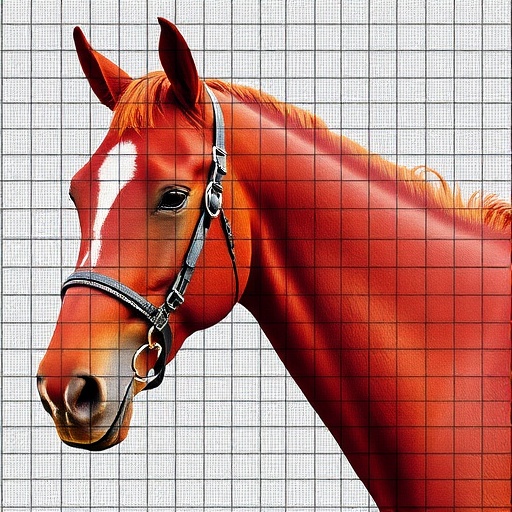
Image Credits: AI Generated
DOI: 10.1093/jvimsj/aalag080
Keywords: equine cardiology, cardiac arrhythmias, 12-lead ECG, vectorcardiography, premature depolarizations, dynamic time warping, catheter ablation, sudden cardiac death, horses
Cite Scienmag News
APA MLA Chicago
William Thompson. (September 3, 2026). Using 12-lead ECG to locate premature heartbeats in horses. Scienmag. https://scienmag.com/using-12-lead-ecg-to-locate-premature-heartbeats-in-horses/
Copy citation Download RIS
Tags: 12-lead ECG accuracy in horses12-lead ECG in horsescardiac diagnostics in horsesdiagnosing arrhythmia origin in horsesECG interpretation for horse arrhythmiasequine arrhythmia diagnosisequine cardiology advancementsequine cardiovascular disease managementequine electrocardiographyequine ventricular arrhythmia detectionlocalization of premature heartbeats in equineslocation of premature heartbeats in horsesnoninvasive cardiac diagnostics in horsesnoninvasive cardiac monitoring in horsessudden cardiac death risk assessment in horsestargeted treatment of equine heart arrhythmiastargeted treatment of horse cardiac arrhythmiasvectorcardiography for horse arrhythmiasvectorcardiography for horsesventricular arrhythmia localization in horsesveterinary internal medicine cardiac studies